
They ruled this planet for at least 3 billion years before the evolution
of eukaryotic life forms. Their descendants are within our cells as the
mitochondria and in plants as the chloroplasts.
"Adam had 'em"
| 1670's Anton von Leeuwenhoek | first to see microorganisms and record the information; verified by Royal Society |
| 1830 Joseph J. Lister | the father of Joseph (Lord) Lister published his work on improving the quality of microscopes - a major advance in the use of the microscope for visualization of microbes |
| 1877 Robert Koch | develops methods for staining and photographing bacteria |
| 1878 Ernst Abbe | describes oil immersion lens |
| 1881 Paul Ehrlich | introduces dye methylene blue into bacteriological staining |
| 1881 Hans Christian Gram | describes a new method for staining bacteria - the Gram stain (a differential stain) |
| 1881 Robert Koch | publishes his methods for the isolation of pure cultures by the use of semi-solid media, firmly establishing bacterio- logy as a science |
| 1887 R.J. Petri | describes new type of culture dish for semi-solid media |
| 1911 Oskar Heimstadt | invents fluorescent microscope |
| 1932 Max Knoll &Ernst Ruska | publish the first description of the transmission electron microscope (TEM) |
| 1935 Frits Zernike | publishes first description of the phase-contrast microscope |
| 1877 Robert Koch | Bacillus anthracis (anthrax) |
| 1879 Albert Neisser | Neisseria gonorrhea |
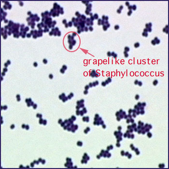
| 1881 Alexander Ogston | Staphylococcus aureus (pyogenic infections) |
| 1882 Carl Gessard | Pseudomonas aeruginosa (various) |
| 1882 Robert Koch | Mycobacterium tuberculosis |
| 1882 Frederick Fehleisen | Streptococcus pyogenes |
| 1883 Theodor Klebs | Corynebacterium diphtheriae |
| 1884 Friedrich Loeffler | Corynebacterium diphtheriae |
| 1884 Arthur Nicolaier | Clostridium tetani (anaerobe) |
| 1884 Robert Koch | Vibrio cholerae |
| 1884 George Gaffky | Salmonella typhi |
| 1885 Gustav Hauser | Proteus vulgaris (various) |
| 1885 Theodor Escherich | Escherichia coli (normal flora) |
| 1886 Daniel Salmon & Theobald Smith | Salmonella cholerae-suis (swine plague) |
| 1887 David Bruce | Brucella melitensis (brucellosis) |
| 1888 August Gaertner | Salmonella enteritidis (food poisoning) |
| 1889 Shibasaburo Kitasato | Clostridium tetani |
| 1892 William Welch & George Nuttall | Clostridium perfringens (gas gangrene) |
| 1894 Alexandre Yersin | Yersinia Pasteurella pestis (bubonic plague) |
| 1897 Emile van Ermengem | Clostridium botulinum |
| 1898 Kiyoshi Shiga | Shigella flexneri (bacterial dysentery) |
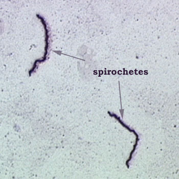
| 1905 Fritz Schaudinn & Erich Haffman | Treponema pallidum (syphilis) |
| 1906 Jules Bordet & Octave Gengou | Bordetella pertussis (whooping cough) |
| 1909 Howard T. Ricketts | Rickettsia rickettsii (Spotted Fever) |
| 1912 Hideyo Noguchi | Spirochaeta refringens (first pure culture of a spirochete) |
| 1912 George W. McCoy & Charles W. Chapin | Pasteurella tularensis (rabbit fever) (rediscovered and renamed Franciscella tularensis in the 1940's) |
The basic idea of immunology was known to the early Greeks and recorded by the historian Theophrastus as he observed soldiers dying or recovering from both wounds and other infections.
The term "immune" was first applied to those soldiers who survived the Black Death (bubonic plague) and were therefore immune from battle field service. It was their chore to cart off and burn the dead bodies of plague victims. A tough way to start as the first male nurses.
| 1660's William Harvey discovers that blood circulates in a closed system of arteries, capillaries, and veins - thus disproving the early ideas of the Roman physician Galen |
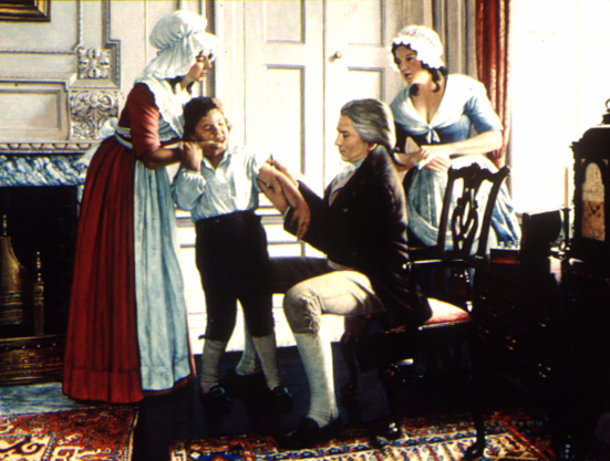
| 1798 Edward Jenner, an English country doctor, develops a vaccine for smallpox and initiates attenuation studies |
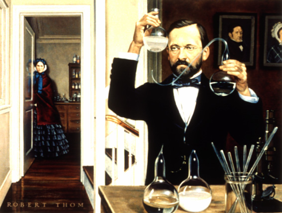
| 1882 Pasteur, using some of Jenner's ideas, attenuates the agent responsible for fowl cholera and goes on to develop vaccines for anthrax and rabies |
This work represents the beginning of what is called active immunization. |
| 1884 Metchnikoff, a Russian scientist, describes "phagocytosis" by white blood cells (neutrophils); this work establishes a cellular aspect of immunology; Metchnikoff joins Pasteur |
| 1884 Nuttal demonstrates the bactericidal action of serum: Bordet who later discovers the causative agent of whooping cough, explains the serum's action is due to special serum proteins called "antibodies"and other proteins with enzyme-like qualities called complement |
| 1888 Roux and Yersin, both of whom work with Pasteur, isolate and describe the toxin produced by the diphtheria microbe |
| 1890 von Behring and Kitasato isolate and describe the toxin produced by the tetanus organism |
This work sets the stage for the development of passive immunization techniques. |
| 1906 Wasserman adapts the "complement-fixation" reaction for use in syphilis testing |
| 1930 Frobisher and Davis use the complement-fixation test for yellow fever testing |
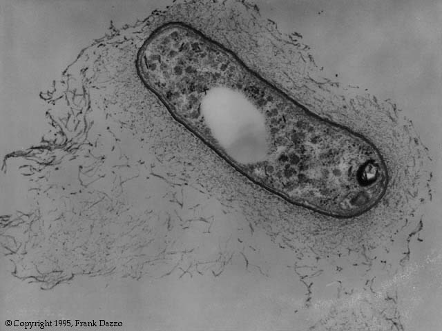
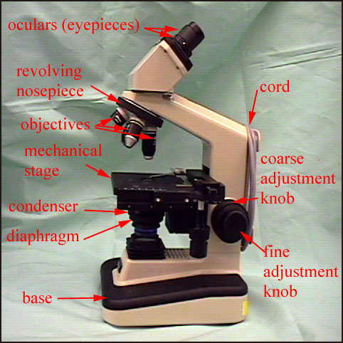
The initial development of microbiology would not have been possible without
the ability to see the microbes. From the time of Leeuwenhoek and earlier,
crude microscopes were available. In the 20th and 21th centuries the types
of microscopes that have been and will be developed has changed dramatically.
These new microscopes now have the capability to see individual molecules
and even individual atoms.
|
 Back to Homepage
 Back to BIOL 2275 Start Page
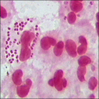
 Note the bacteria on the surface of this white blood cell.
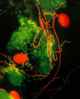