
Introduction
Serious research into the biochemical mechanism of Celiac disease did not begin until the early 1990’s. Prior knowledge regarding the disease came from medical encounters and the interpretations of these encounters from a limited immunological point of view. Doctors found that the disease was common among families and often shortened the life expectancy of the individuals due to the early appearance of colon cancer. Through examination of biopsies from their patients, they were able to determine that there was a connection between the substance gluten and Immunoglobulin A. The patients were then told to simply avoid all products containing gluten, and they often saw a decrease in symptoms and a reversal of intestinal lesions. It was postulated that Celiac patients had genetic sensitivity and predisposition to form an IgA antibody gluten complex. While the early ideas of the mechanism of Celiac were close to understanding the pathogenesis of the disease, it was not until early in the 21st century that true understanding began. Research has been underway at Stanford University in conjunction with several other universities throughout the world since the late 1990’s. The primary finding of this research has been the determination of the mechanism by which Celiac disease proliferates. The mechanism consists of several components including T1 helper cells, gluten epitopes called gliadins, an enzyme called transglutaminase, and histocompatibility leukocyte antigens (DQ2). The primary T1 helper cell is known as CD4 (Figure1), which is an intestinal T cell that is often extracted from the biopsies of patients.
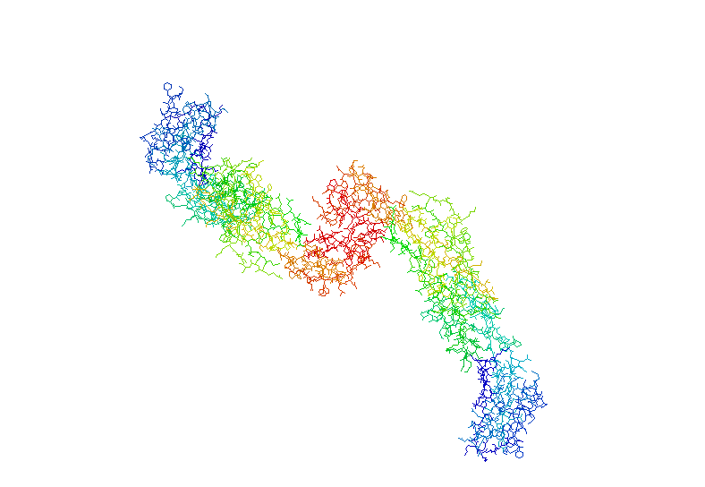
Figure 1. Structure of CD4 T1 helper cell found in the intestinal tract of Celiac disease patients. This T cell recognizes the gluten epitopes as foreign and designates them for destruction
Transglutaminase (Figure 2) is one of the enzymes present in the digestive tract that is responsible for the degradation of actin filaments and more importantly it degrades gluten into the gliadins that catalyze the autoimmune response.
![]()
Figure 2. Structure of transglutaminase, the primary enzyme responsible for the degrading of gluten. The resultant products of this enzymatic reaction are the gliadins that promote the autoimmune response in Celiac disease
Perhaps the most vital portion to the sequence of the mechanism is the binding of the gliadin peptides to the HLA DQ2 (Figure 3), which is the final step before the initiation of the autoimmune response. The HLA has a specific affinity that is much higher for the negatively charged epitopes of gluten than any other substance found in the intestinal tract.
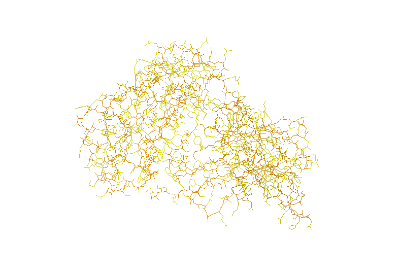
Figure 3. Structure of HLA DQ2. The gliadins bind to the HLA, which then catalyzes the autoimmune response, destroying the intestinal tract
To better understand the mechanism, a series of different methods and materials were used to elucidate the composition of the gliadins, in addition to the structure and function of transglutaminase, T1 helper cells, and HLA DQ2.
To return to the Welcome page click here
Feel free to email me with any questions!!
Sources included on this page:
2,3,4,5 
