|
Objectives:
Introduction
Part I
Eubacteria consist of the bacteria and cyanobacteria. The typical prokaryotic organization lacks membrane-limited organelles such as nucleus, mitochondria, and lysosomes. They have a
Bacterial shape, while genetically determined, is influenced by environmental factors. Shape is round or coccal shape, rod-shaped or bacillus shape or spiral or spirillum shaped. A variety of views are available in the links below. The following link will provide general Information and graphics for the Eubacteria. Use the links below to get a closer view of the organisms.
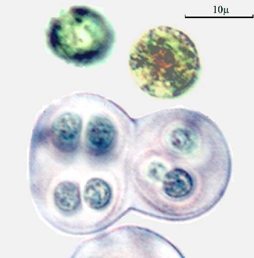
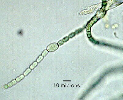
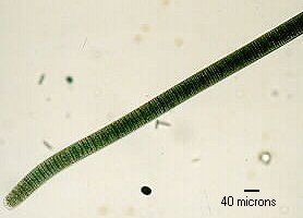
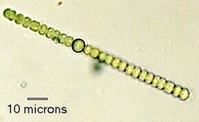
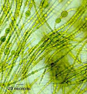
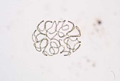
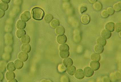
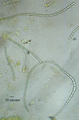
The cyanobacteria, originally called the blue-green algae, are photosynthetic prokaryotes. Their cells are generally larger than the bacteria and are covered or surrounded by a gelatinous covering or sheath. Cyanobacteria come in all the colors of the rainbow and some contain a swollen or enlarged region of specialized cells called a Heterocyst. The heterocyst contains the enzymes necessary to carry out the process of nitrogen fixation where atmospheric N2 is converted into other forms usable by plants.
Use the links below to get a closer view of the organisms.
In the absence of microscopes and slides, use the embedded links below to learn about the various organisms.
See also pages 25-28 in A Photographic Atlas for the Biology Laboratory, 4th Edition.
Part II Sampling Your Environment
If you have access to a school lab and can procure petri dishes with nutrient or tryptic soy agar then please use one of them. If you would like to try being a microbiologist (and cook) at home then follow the instructions below.
Making a Microbiological Medium (used by permission of the developer, Pamela P. Tabery with modification by dse)
You can use a disposable foil muffin tin or the bottom one inch of a 24 oz. water bottle as your petri dish. Either can be covered with aluminum foil to prevent contamination and moisture loss. A very simple medium could be made from unflavored Knox gelatin all by itself but it contains only protein and would not support the growth of many organisms. This will make enough for one culture. Adjust the size of the recipe by multiplying each ingredient and the water by the number of dishes you are attempting to fill. Pour the following into a container and mix: 1 teaspoon (Knox) unflavored gelatin, 1/2 teaspoon sugar, and 1/2 teaspoon instant beef-flavored bouillon. Pour 1/4 cup boiling water into the bowl and mix. Pour this mixture into your muffin tin or substitute petri dish. It should solidify at room temperature. If the solution does not seem to be solidifying in a reasonable time, then add more gelatin next time. Use a cotton Q-tip as your swab. You could sample the microbes on your skin also by swabbing the skin surface or by placing your finger directly on the surface of the medium. Only your imagination limits the types of sampling you could do. Cover the dishes after you have gently swabbed the surface. Swab a dish without a sample on the Q-tip to serve as a control and see what is on the Q-tip. Dispose of the plates by adding a little bleach or Lysol type product to the surface and placing everything in a plastic bag. Compare the types of organisms you find with the areas you sampled.
|