There are more than 100,000 species of identified fungi. Of these, between
50 to 100 are know to be pathogenic to humans. Only about 20 species cause
fatal infections. Diseases caused by fungal agents are called MYCOSES and
can be divided into four major subdivisions based on the site of infection.
These groups are: the SUPERFICIAL, the CUTANEOUS, the SUBCUTANEOUS, and the
SYSTEMIC mycoses. A fifth group is also included. They are known as the
OPPORTUNISTIC mycoses and are seen in a compromised host.*
(see also: The Emerging Fungal Threat, Science, Vol.266, pp. 1632-1634, December 9, 1994)
Asexual Spores Formed by Fungi**
Conidia - This term is sometimes used generically for
all asexual spores. It may be used specifi-
cally for spores borne singly or in clusters
along the sides or at the tips of hyphae or
of specialized hyphal branches called conidi-
ophores. They are highly diversified in shape
size, color and septation. Large (usually
multinucleate) and small usually uninucleate,
conidia are called macro- and microconidia
respectively. Examples of genera forming
this type of spore: Aspergillus, Penicillium,
Cephalosporium, Microsporum, Trichophyton
Arthrospores - These are cylindrical cells formed by a
double septation of hyphae. Individual cells
called spores are released by fragmentation.
Examples: Coccidioides, Trichophyton
Blastospores - Buds that arise from yeast or yeast-like
cells. Examples: Candida, Saccharomyces
Chlamydospores - Thick-walled, round spores formed from
terminal or intercalated hyphal cells.
Examples: Candida
Sporangiospores - Spores formed within sac-like structures
called sporangia at the end of hyphal stalks
or on special hyphal branches called sporan-
giophores. Examples: Coccidioides, Rhizopus
1. The Dermatophytes (Cutaneous Mycoses)
The dermatophytes are a closely related group of fungi which cause specific
infections of humans and animals. The diseases they cause are referred to as
DERMATOPHYTOSES, RINGWORM, or TINEA. They invade only the superficial
keratinized areas of the body such as the skin, hair, and nails. They do
not cause systemic infections and rarely, if ever, invade the subcutaneous
tissues.
Cutaneous mycoses represent the most common fungal diseases in humans and
are important health problems in countries where over-crowding and lack of
simple hygiene exist. There are three genera of dermatophytes that cause
infection: Trichophyton, Microsporum, Epidermophyton. Cutaneous
diseases can be treated by oral admini-stration of Griseofulvin, an
antibiotic which has an affinity for keratinized tissues. Topical ointments
such as Tolnaftate (Tinactin), Haloprogrin, and Miconazole
are effective but require prolonged administration. Other ointments
sometimes used are Desenex and Whitfield's.
a. Genus Trichophyton
Microscopically, microconidia are the prominent spore forms. Various species
are responsible for ringworm of the scalp (tinea capitis) and body (tinea
corporis). Some species are common causes of "athlete's foot"
(tinea pedis). Other species may cause tinea unguium, tinea barbae, and
tinea cruris. The hair and nails may also be invaded.
b. Genus Epidermophyton
Microscopically, only broad to oval macroconidia are produced. This genus
is represented by a single species, E. floccosum, and is found only
in man. It grows in epidermis (especially in intertriginous areas, as
between the toes), but the hair is not invaded. It is usually responsible
for either tinea cruris or tinea pedis.
c. Genus Microsporum
Microscopically, large, spindle-shaped, multi-celled, rough, thick-walled
macroconidial spores are numerous and characteristic. Most species infect
the hair of children, domesticated and wild animals. Children commonly
acquire the infection from dogs and cats. One specie, M. audouini,
used to be the most frequent cause of ringworm of the scalp in children in
the U.S. but now shares equal billing with Trichophyton tonsurans.
The genus may cause tinea corporis, or tinea capitis.
2. Subcutaneous Mycoses
Subcutaneous mycotic infections are usually initiated by penetration of the
skin with contaminated splinters, thorns, or soil. Once established, these
infections tend to remain localized in subcutaneous tissues and tend to be
extremely persistent. Treatment may sometimes require surgery plus
antifungal agents such as Amphotericin-B. Potassium iodide may be
used topically.
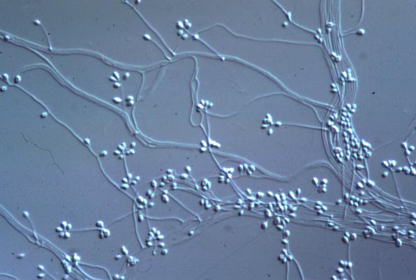
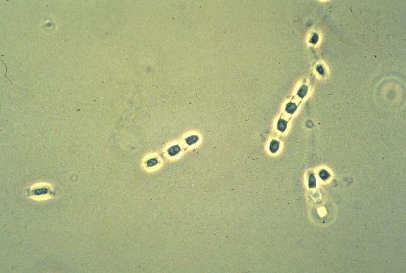
a. Sporothrix schenkii
This organism is single-celled, cigar-shaped and Gram +. It is found within
macrophages or polymorphonuclear cells of lesions or exudates from humans
and animals. The disease it causes is known as sporotrichosis. The disease
is characterized by an ulcerated lesion at the site of inoculation. Multiple
nodules and abscesses occur along the superficial draining lymphatics. The
disease is usually sporadic among farmers and gardeners. A few industrial
outbreaks have occurred among workers exposed to batches of heavily
infected timbers or plants. The spores can also be inhaled. The organism is
found in North, Central, and South America as well as Africa. In the United
States, it is found along the Mississippi and Missouri valleys and
throughout the Northeast.
b. Hormodendrum (Fonsecaea) pedrosoi
This fungus, with at least two others, Hormodendrum F. compacta and
Phialophora verrucosa, produce the disease known as
chromoblastomycosis. Infection arises from penetration of the skin by
contaminated splinters or soil. The infection is most common in the tropics
although its distribution is worldwide. The infection is seen on the legs
of bare-legged laborers and lesions appear as warty, ulcerating,
cauliflower-like growths.
3. Systemic Mycoses (Diseases Involving the Internal Organs)
These infections can penetrate the epithelial tissues and become
disseminated throughout the body. Infections arise most frequently from
inhalation of spores found in the soil. If the infective dose is sufficient,
disease can result. Infection usually starts by development of lung lesions
which may resolve themselves with no further damage. Some diseases may
become chronic and the symptoms often mimic tuberculosis. If the fungal
agents become disseminated into the bloodstream, other internal organs may
become infected.
The treatment of systemic mycoses include Amphotericin-B alone or in
combination with 5-fluorocytosine. More recently Ketoconazole
has shown significant control and relief.
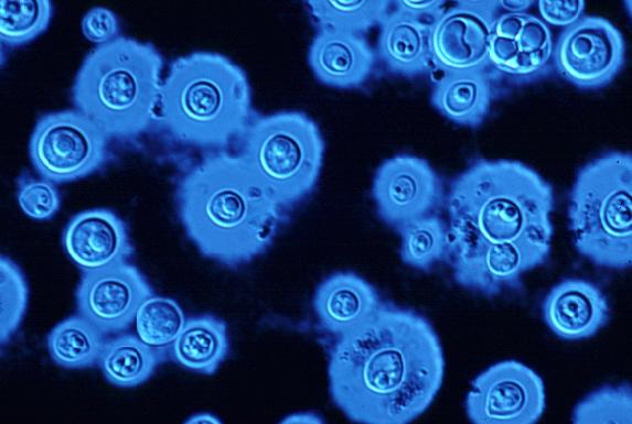
a. Cryptococcus neoformans
This yeast-like, non-sporulating, non-mycelial, budding fungus is
characterized by the development of a wide capsule in tissue and culture.
It has an attraction for the central nervous system and is often considered
the most dangerous of the systemic mycoses. The disease is called
cryptococcosis. Inhalation of the cells is assumed to initiate pulmonary
infection, with subsequent spread to other viscera and the CNS by way of
the bloodstream. Minor infections are common. Amphotericin-B is
usually effective.
In the severe, chronic, and disseminated form of the disease, the brain,
meninges, lungs, and other viscera, skin, and bones are involved to varying
extents in different patients. Chronic meningitis is the most frequent and
mimics tubercular meningitis. The lesions may simulate brain abscesses or
brain tumors. Pulmonary lesions are usually inapparent. The disease appears
sporadically and in essentially all parts of the world. The organism has
been isolated from soil, particularly when enriched with pigeon droppings.
Since the fungus remains viable in dried materials for many months,
contaminated materials are a potent source of airborne infections. The
organism is also opportunistic in the compromised patient.
b. Blastomyces dermatitidis
This organism is spherical, thick-walled, budding, and yeast-like in
tissues, exudates, and cultures at 370 C. It produces a granular
infection of the skin and internal organs which appears very similar
clinically and histologically to tuberculosis. Infection apparently begins
in the lungs and spreads, by means of the circulatory and lymphatic systems,
to the bones, skin, prostate (in males) and other viscera. The
gastrointestinal tract is normally spared. The skin lesions are often quite
conspicuous. The disease is largely confined to Canada and the United
States, particularly in the Mississippi Valley and east to the Carolinas.
Once again, pigeon droppings provide a rich growth medium.
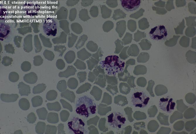
c. Histoplasma capsulatum
This organism is a small, oval, yeast-like fungus in tissues and cultures.
In infected tissues, it is usually localized in macrophages and reticulo-
endothelial cells. The organism is present in soil and inhalation of spores
leads to pulmonary infection. Miliary (seedlike) nodules and lesions appear
through-out the lung parenchyma and hilar lymph nodes become enlarge. The
initial infection is mild and may pass unnoticed. In a small number of
infected individuals the infection becomes progressive and widely
disseminated, with lesions in practically all tissues and organs. Fever,
wasting, and enlargement of liver, spleen, and lymph nodes occur and the
disease may closely simulate tuberculosis. The disease is known as
histoplasmosis or Darling's disease. In some parts of the country it is so
prevalent that it is called "summer flu." It is localized in
areas that have been enriched with bird excreta, especially from birds such
as starlings, chickens, crows (but not pigeons), as well as bats. It is
endemic in the Mississippi River Valley and it is estimated that over
30 million people in the U.S. have been infected.
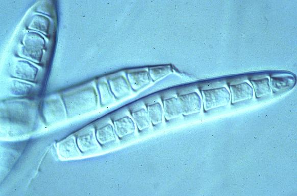
d. Coccidioides immitis
A spherical, thick-walled endospore filled organism occurs in tissue and
exudates. The disease, known as coccidioidomycosis, is highly infectious
as arthrospores are easily spread. It is the most virulent of the systemic
mycoses. It may produce an acute, benign, primary, self-limited respiratory
infection or a chronic, malignant, secondary, progressive, disseminated
infection usually referred to coccidioidal granuloma. The organism grows as
a saprobe in desert soils of the southwestern United States and northern
Mexico. It is sometimes known as Valley Fever. Infection is established by
inhalation of airborne spores.
Of the 52 reportable diseases, this is the only fungal disease organism that
is reportable.
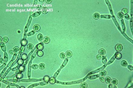
e. Candida albicans
This oval, budding, yeast-like fungus produces blastospores and pseudo-
mycelia in tissues and exudates. Its exact etiologic significance in any
disease process is difficult to establish since it is often present in the
mouth and intestinal tract of healthy individuals. It is often a secondary
contaminant in other recognized diseases. It may truly be considered an
opportunistic organism because it may cause cutaneous, subcutaneous,
mucocutaneous, or systemic infections. When Candida becomes invasive
it establishes a variety of acute or chronic, localized or widely
disseminated lesions. Dependent upon body site infected, Nystatin or
Candicidin may be used as well as previously mentioned drugs.
A number of fungi are not pathogenic in healthy humans, but may become
virulent pathogens in those suffering from a variety of disorders, and in
those treated intensively with broad-spectrum antibacterial drugs or with
immunosuppressive measures.
Fungal diseases are the exception and not the rule. The fungal agent often
induces a cell-mediated immune response in which there is inflammation and
a walling off of the agent in a fibrous, calcified deposit.
The two most important factors that determine ones susceptibility to disease
are the number of organisms to which the host is exposed (infective dose)
and the general state of health of the host at the time of exposure.
Allergic reactions in some individuals are not surprising since spores are
frequently inhaled.
* modified from General Microbiology, Boyd, p. 691
** modified from Microbiology, Davis, Dulbecco, et.al., p. 972
summary chart - Fundamentals of Microbiology, Alcamo, pp. 456-457

 Back to Homepage
 Back to Menu Page
 Back to Microbiology Start Page