GRAPHIC WARNING
Examination is completed for Pygocentrus nattereri (Paraguay/Argentina region) commonly known as "ternetzi"
May 23, 2004
History of Fish: A summarized detail about death
Only had the fish 1 (no more than 2 weeks) before its death. First came in the
top part of the head was really thing (like caved in), other marks were a
healing wound (probably ammonia) burn on the right body behind the gills (should
still be present since it hasnt healed totally), and close to tail fins. Also
when fish was housed in the tank, it never just took a spot to call his own, but
usually swam mid center of the tank, mostly in circles and not the usual end to
end. Diets consist of feeders, mostly beef heart, and 1 mice that was
dared to try. He was the greediest eater in the tank, and never took shibby from
the Alpha of the tank (another 13" Tern).
3 days prior to his death, both eyes weren't aligned, a bit sunked in,
when looking directly face to face. 2nd day, eyes were really sunked in upward,
to the point where the eyes weren't present and just empty eye sockets. Tank
perimeters PH 7.0, 0 ammonia, Nitrite .5 Used a hospt tank with same water but
added new to keep the P from stressing. Left 5 feeders for him to try and eat
(which non were touched), and confined him in a covered tank to ease him from
the light. Checked his status in the morning.. alive but looked immobile,
breathing slow and steady. Came home from work 8 hrs later, Tern was dead..
floating sideways on top of the tank.
|
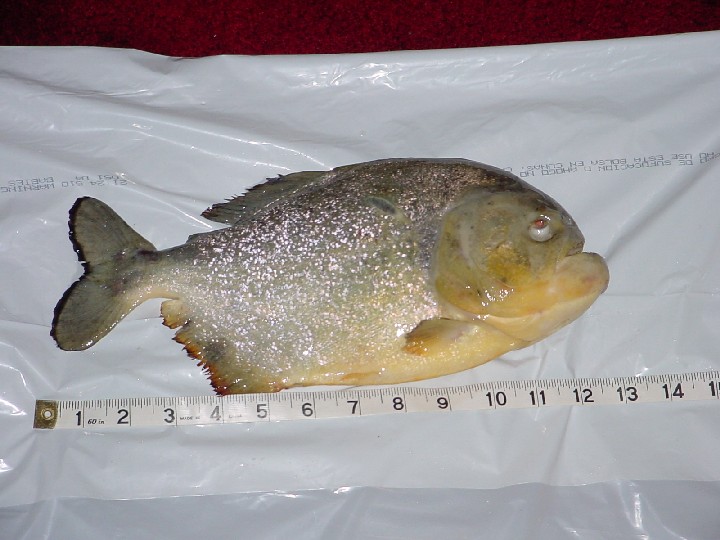
Sex of fish: Male Measurements are metric: SL: 26.5 cm TL: 29.1 cm SW (girth): 13.4 cm Weight: 17.5 oz
Prior to Death, then death: |
|
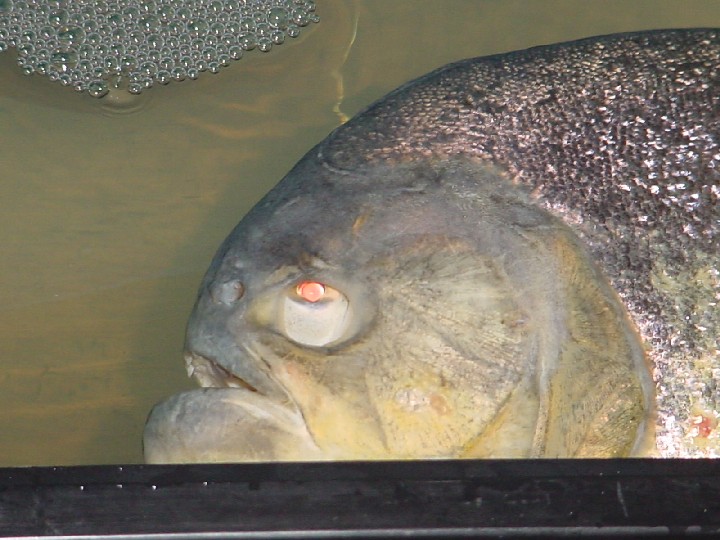
Appearance of specimen (External)
The fish has been dead for (approximately) since May 18, 2004. It was received here on May 22, 2004. It was packed in a cloth, soaked in isophrophyl, placed in 4 plastic bags (zip lock). This was then sealed in popcorn plastic shipping material.
Removing the seal revealed a fish that was of good size. The belly appeared distended. Eyes were sunken in as if shoved in. Iris of eyes appeared distorted and fixed. Holes were noted on the opercle and head area. In these photos (above) one can see after death the fish shows signs of systemic mycobacteriosis. The chronic ulcers are seen on the flank and on the opercle. This disease is caused by a number of different bacterial species of the genus Mycobacterium. No external parasites were seen. Scraping and view under microscope revealed nothing of interest. 99% probability parasites vacated host at or last stages of death.
Internal exam
Post mortem exam reveal (see photos below) gray to white nodules (granulomas) on the internal organs, primarily the spleen and kidney. These problems are an indication of a lack of Vitamin; C, E, and B-complex. Gills (portion) are removed and are milky, scrapings reveal debries, placed on slides. Under microscope no discernable parasites, Uninteresting.
The fish appears pregnant, but in opening the fish, I found an inflated air bladder, and gonads were also inflated. This indicates blockage of some type. Removal and incision made of both reveals nothing interesting. It is not unusual that air bladder remains inflated at mortality. The thickness of the bladder indicates bacterial infection. The gonads may likely remained inflated from fishes last breath. Not unusual and no parasites seen either visually or under microscope. Swelling of the abdominal cavity is part of the disease phase of "dropsy".
Ulceration was noted in nearly all organs consistent with mycobacterial infection, a gram-positive bacteria. I reversed the organs to look at both sides, ulceration was present in spots.
Acites (bloating) present with exophtalmia (popeye) which I believe is not indicative here, shows signs of damage to kidneys and liver. The scales around the belly area down to the serrae reveals protruding scales. Specimen had to be raised from the flat surface in order to better see this scale postions. This does indeed indicate fluid under them. The eyes are sunken (endopthalmia) and is consistent with bacterial infection, probably attributable to "dropsy", a common diseases of fishes.
This
diseases is normally spread from infected fish that are either sick or dying and
it is eaten by the predator fish. According the hobbyist, live feeders
were part of this fishes diet. Goldfish are said to be somewhat more prone to
dropsy than other fishes. In some cases it is caused by a Costia
Infection. Unknown what condition feeders were in.
The most common cause of dropsy is an internal bacterial infection, while
viruses and poor water quality have also been associated. The condition affects
the fish's internal organs, and as a result they cease to function correctly. A
concentration of fluid in the body tissues and cavities causes the fish's
abdomen to become swollen and appear bloated. No such fluid was detected in the
fishes cavity, but this is simply one symptom and may not be present in this
mortality. All things being equal based on this exam indicates that dropsy is
indicative.
No tapeworms detected.
DIAGNOSIS
I'm considering that the problem is probably a gram-positive bacterial problem that was probably transfected by feeders. It's quite probable that lack of suitable vitamins (common with ornamental fish care by hobbyists) may have helped cause further deterioration of the fish to resist disease. I do not believe the conditions are attributable to tuberculosis as the fish doesn't show any outward signs of this problem. So this is ruled out as a possible condition. All physical anomolies are attributable to dropsy and are symptomatic of the disease.
1. Blood Flagellates - Normally aquarium fish are not affected by this. This disease consists of single-celled organisms with hair-like flagellae. They live as parasites in the blood of the fish. They live in a leeches intestines and are passed on to the fish when the leech bites the fish. Symptoms: Fish will appear listless and swim abnormally. They become emancipated, with sunken eyes and pale gills (this indicates low red blood cells). Severely infected fish will die. This condition does not appear to have been the cause. So this is ruled out. One should always consider that feeder fish in farms are susceptible to leeches and while the probability is there that the goldfish might have been infected, it remains unknown for this exam.
2. Types of dropsy -
a) Acute Dropsy - Internal bacterial infection can cause internal bleeding and thus cause acute dropsy.
b) Chronic Dropsy - * cancer: In this case, the abdomen is slow to swell as the cancer affects the fish's internal organs. If the fish is not isolated in the early stages of the disease, it could spread to other fish that are being housed with the ill fish. Chronic Dropsy - parasites: Internal parasites can cause dropsy(abdominal swelling) because they are rather large parasites or because of the damage they are causing with the fish's organs. The abdomen tends to swell over a period of time if the fish is infested with internal parasites. It is best to isolate the sick fish at once to help maintain the outbreak of disease with other fish.
Treatment
Dropsy is a fatal disease and communicable. Fishes infected should be isolated immediately. There is no cure for dropsy. A Google search revealed suggestions on medications, but it appears the disease is untreatable and the only preventative treatment appears to be vitamins (These problems are an indication of a lack of Vitamin; C, E, and B-complex) and good water conditions. It is my suggestion/recommendation that live feeders be removed as a source of food, particularly if they are NOT closely watched for outward disease/parasite problems before being fed to the predator. Vitamins should be fed to the remaining fishes in the main fish tank. Those fishes should be closely monitored for any outward signs of distress or morphological signs of unusuality; frayed fins, discoloration, ulcerations, cloudiness of eyes. These should all be treated with medications, preferably wide spectrum. Beefheart if fed on a regular bases should be free of fat and veins. It's probably this fish suffered from cancer of the intestines.
|
|
|
Completed May 23, Sunday
USE YOUR BACKSPACE TO RETURN RESEARCH
TO RETURN HOME CLICK HERE.
The OPEFE web site and its contents; is disclaimed for purposes of Zoological Nomenclature in accordance with the International Code of Zoological Nomenclature, Fourth Edition, Article 8.3 and 8.4. No new names or nomenclature changes are available from statements at this web site.
Copyright© 1994-2012 Oregon Piranha Exotic Fish Exhibit (The OPEFE fish exhibit is permanently CLOSED as of 2000) Sutherlin, Oregon. Information posted on this web site is archival data on fish scientific classifications and other information. DISCLAIMER: The copyrighted material may not be used for any purpose other than private study, scholarship or research. Cited information requires credit and this link www.opefe.com. All rights reserved. All images shown (unless otherwise noted) is property of OPEFE.