
New UV emitters : Radioisotopes and XRF sources explained
by first mapping of photon , electron, proton, and neutron
M.A. PADMANABHA RAO
114 Charak Sadan,
Vikas Puri,
Abstract. The `UV dominant atomic spectra from solids' newly detected at room temperature from salts or metals present as XRF sources and radioisotopes like 137Cs (radiochemical), and 57Co (cobalt metal) represent fundamental optical emission caused by previously unknown core-valence excitation. Spectacular 28 UV photons caused by each of the 4000 Rb X-rays emitted from gamma excited Rubidium salt (Rb XRF source) is noteworthy. The new physical insight, same performance for gamma, X-ray, and beta at any given energy during Coulomb interaction, in contradiction to beta's known particle behavior could be due to the fact that excited atom recognizes their energy as the sole criterion. To resolve such key issues in physics, the author attempted in first ever mapping of photon, electron, proton and neutron through advanced Yoga technique. The sketches of proton, electron and gamma photon revealed that they all possess electric, magnetic, and gravitational fields providing them a shape and size. Increase in shape and size of these fields particularly for an electron when escaped from atom better explains why electron behaves like photon within excited atom and as a particle outside. Reduction in size of electron and photon with increase in energy explains why gamma reaches faster than light photons from gamma- ray bursters (GRBs). Electron transforming into gamma or X-ray and vice versa in Coulomb field seemingly a different manifestations of energy. Core electron hooked to proton, better explains its rotation in fixed orbit, could be one step ahead to Bohr's atom.
Key words: radioisotopes; XRF sources; UV,VIS, NIR emissions; Bharat radiation; atomic phenomenon; room temperature atomic spectra; metallic solids; Core- valence excitation.
1. Introduction
Previously unexplored area of subatomic research into highly excited atoms of solid radioisotopes and XRF sources leading to discovery of their fundamental UV dominant optical emission is the highlight of the current paper. Spectacularly high counts detected from unexpected Rb XRF source (AMC 2084, U.K.) unusually kept on bare PMT (9635QB, THORN EMI) hinted the possibility of optical radiation when steeply dipped on keeping a thin black polyethylene sheet in between source and PMT demanding confirmation by a foolproof method.
Despite the limitation that these calibration
sources yield very poor light intensity that cannot be seen, use of a pair of
sheet polarizers enabled characterization of the optical spectrum from UV (up to
400 nm), VIS (400 to 710 nm), and NIR (beyond 710 nm) radiation intensity
measurements. Two extraordinary spectral features: (1) UV dominance and (2)
dependence of UV,
The unprecedented UV dominant optical radiation detected from 57Co metallic solid provided definite clue on atomic emission by free metal atoms, formed as a result of valence excitation of nuclear and, or core excited atoms situated in between unexcited atoms (Padmanabha Rao 1997, 1998, 1999, 2001, 2002). The new insight is strongly suggestive of existence of a kind of 'atomic state of matter' within solid radioisotopes and XRF sources at room temperature. The first `room temperature atomic spectra detected from solids' by non-thermal valence excitation could be spectacular advancement in the field of atomic spectroscopy. The author’s explanation that ionizing radiations passing through core - Coulomb field first generate exciting energies at eV level (termed Bharat radiation) necessary to cause valence excitation that in turn generate the optical spectrum within the excited atom is increasingly gaining ground (Padmanabha Rao 1997, 1998, 1999, 2001, 2002).
New understanding of fundamental properties of beta, not exhibiting its particle behavior to be distinctly different from gamma or X-ray stems from the finding, same percentage of UV, VIS and NIR intensities at any given ionizing radiation energy, as has been explained due to their common transit through core - Coulomb field within excited atom in vacuum.
2. Experimental
The radioisotopes selected for the study were present either as radiochemicals or metallic solids in kBq or MBq activities. The handy Variable Energy X-ray source (AMC 2084, U.K.) provided Rb, Ba and Tb XRF from solid Rb, Ba and Tb salts and Cu, Mo or Ag XRF from Cu, Mo and Ag metallic solids during gamma excitation from 241Am.
The experimental set up is nothing but a simple Gamma ray Spectrometer with a difference in its probe (Dinesh Bohra et.al 1992, Padmanabha Rao 1997,1999). Unexpected detection of UV dominant optical radiation did owe its success to (a) the use of bare photo multiplier tube (9635QB THORN EMI) on which the source was directly kept, (b) gain of the linear amplifier set relatively higher than the requirement for Gamma ray Spectrometer, and the time constant at 0.1 msec. Optimally low background rate (13 cps) of the PMT noticed, despite high gain setting ensured satisfactory operating condition of the PMT free from any light leak. Ultimately, 8K MCA displayed a single pulse height spectrum for simultaneously detected optical and ionizing radiation intensities by thin quartz window of the PMT. Accrued the integral counts under the spectrum that represent combined radiation intensity for four minutes but shown in terms of counts per sec (cps) in Table 1.
The parallel pair of diachronic visible light
linear polarizers employed in the study block characteristic UV radiation up to
400 nm but transmit low percent linear polarized visible
(

Figure 1
- Schematic of the author's technique discloses first ever evidence of UV
dominant optical emission from 137Cs. a, When 137Cs source
was kept directly on the thin quartz window of the PMT (9635QB, THORN EMI), the
PMT detected 9098 (6.2) cps presumably due to UV (up to 400 nm),
UV radiation intensity = 8305 (9.8) cps.
VIS radiation intensity = 73 (7.1) cps.
NIR radiation intensity = 201 (6.4) cps.
These intensity estimates disclose first and definite evidence for the UV dominant optical radiation from 137Cs. Unification of data, expressing the estimated UV, VIS and NIR radiation intensities as percent of the gross light intensity could address the problem of unequal source strengths and the standard units of radioisotopes, and XRF sources. Quantitatively, dominant UV from 137Cs has been a staggering 96.81% of the gross light intensity, in comparison to VIS and NIR radiation intensities limited to 0.85% and 2.34% respectively (Table 1).
3. Results and discussion
Detection of analogous UV dominant optical radiation from all the tested XRF sources and radioisotopes has been a key success of the experimental study (Padmanabha Rao 1997, 1999), when previous researchers could neither predict such a possibility nor were able to see UV because of invisibility (Table 1). Radioisotopes like 137Cs, 131I, 137Cs, beta emitter like 90Sr, and Rb XRF source can serve as handy room temperature UV sources in material science. The percent UV, VIS, and NIR intensities of any source showing a clear link with ionizing radiation energy of abundant radiation, regardless of type of emission and source is another major success of the study.
Table1- First evidence on radioisotopes and XRF sources as UV emitters
|
Source |
Emission |
Energy (MeV) |
gross light intensity (cps) |
%UV |
%VIS
|
%NIR
|
|
55Fe |
Mn X-rays |
0.00589 |
125(0.9) |
|
|
|
|
Rb XRF |
Rb X-rays |
0.01339 |
125,321(23) |
99.62 |
0.37 |
0.01 |
|
133Ba |
Cs X-rays |
0.03097 |
2,803(3.9) |
97.51 |
1.46 |
1.03 |
|
Ba XRF |
Ba X-rays |
0.03219 |
2,064(7.3) |
95.64 |
3.83 |
0.53 |
|
152Eu |
Sm X-rays |
0.04012 |
3,052(6.2) |
90.33 |
5.90 |
3.77 |
|
Tb XRF |
Tb X-rays |
0.04447 |
37(1.1) |
|
|
|
|
241Am |
g |
0.05954 |
1678(2.1) |
98.03 |
1.91 |
0.06 |
|
201TI |
Hg X-rays |
0.07082 |
1,830(2.8) |
95.73 |
3.83 |
0.44 |
|
57Co |
g |
0.122 |
626(1.8) |
96.01 |
1.76 |
2.23 |
|
99mTc |
g |
0.141 |
468(4.5) |
94.02 |
3.85 |
2.13 |
|
147Pm |
β |
0.224 |
3,606(3.9) |
99.36 |
0.58 |
0.06 |
|
45Ca |
β |
0.252 |
2,333(3.2) |
95.76 |
4.20 |
0.04 |
|
113Sn |
g |
0.393 |
91,105(23) |
96.95 |
2.21 |
0.84 |
|
141Ce |
β |
0.444 |
727(0.9) |
98.76 |
1.10 |
0.14 |
|
22Na |
g |
0.511 |
2,284(3.5) |
94.92 |
2.50 |
2.58 |
|
137Cs |
β |
0.514 |
8,579(7.0) |
96.81 |
0.85 |
2.34 |
|
131I |
β |
0.607 |
234,079(5.0) |
96.64 |
3.23 |
0.14 |
|
110mAg |
g |
0.6577 |
48,393(28) |
88.07 |
4.36 |
7.57 |
|
204TI |
β |
0.763 |
84,984(24) |
96.60 |
2.96 |
0.44 |
|
59Fe |
g |
1.099 |
39,985(16) |
95.98 |
1.52 |
2.50 |
|
60Co |
g |
1.33 |
2,207(3.4) |
92.98 |
2.31 |
4.71 |
|
86Rb |
β |
1.77 |
38,677(23) |
73.56 |
9.82 |
16.62 |
|
90Y |
β |
2.27 |
29,563(19) |
83.36 |
8.02 |
8.62 |
|
Sources present as metallic solid | ||||||
|
Cu XRF |
Cu X-rays |
0.00805 |
22(0.8) |
|
|
|
|
Mo XRF |
Mo X-rays |
0.01748 |
27(0.9) |
|
|
|
|
Ag XRF |
Ag X-rays |
0.02216 |
30(1.0) |
|
|
|
|
57Co |
g |
0.122 |
6,343(8.1) |
88.18 |
5.71 |
6.11 |
|
60Co |
g |
1.33 |
30,123(34) |
|
|
|
The number in the parenthesis denotes standard deviation of counts. The counts caused by ionizing radiations are intentionally not shown here, light emission being the main focus of the study.
Gross light intensity measurements made from Cu, Mo, and Ag XRF sources, and 60Co metallic solid, and UV dominant spectrum observed from 57Co metallic solid provide the first ever evidence of metallic solids giving rise to room temperature optical radiation (Table 1). Rb XRF source exhibiting excellent optical properties may have significance in material science. That the observation 4400 Rb X-rays in single 'pi' geometry causing high quantum yield, 125,321(23) UV photons in one sec from Rb XRF source analogous to strong lines in Rb atomic spectrum provided strong evidence on atomic emission (Padmanabha Rao 1997, 1999).
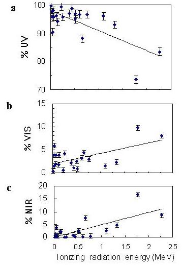
Figure 2. (a) Unprecedented UV dominance, and (a, b, c) key control of energy of ionizing radiation with maximum abundance on percent UV (up to 400 nm), VIS (400 to 710 nm), and NIR (beyond 710 nm) radiation intensities as shown in Table 1 have been the two emissive features of the new spectra commonly observed from both XRF sources and radioisotopes.
The current study may hopefully prompt futuristic line spectra with highly active soft beta emitters like 90Sr on exposing the PMT for long hours. The current study could practically be a laboratory model for the well established astronomical phenomenon of brilliant after glow at optical wavelengths from X-ray Pulsars and Gamma ray Bursts (GRBs).
Eventually, the current subatomic research enabled
new understanding of fundamental dynamical properties of beta, gamma, and X-ray
while they make a common transit through core – Coulomb field in vacuum within
the core- excited atoms, inaccessible outside excited atoms in any medium
(Padmanabha Rao 1997, 1998, 1999, 2001, 2002). Fig 2 provides experimental
evidence on same percentages of UV,
To better understand the fundamental properties of photon, electron, proton, and neutron, their sketches were drawn through advanced Indian yogic technique. For the first time, their shape could be known by mapping when not previously possible to scientists by any other means. The sketches reveal previously unknown gravitational field to photon, electron, proton, and neutron (fig.3). Proton has shown spin (fig.3, bottom left). Interestingly, electron is attached at the centre of proton retaining their individual identity, while acting together as neutron (fig.3, bottom right). As a result of attachment, proton lost its characteristic spin.

Figure 3. First ever sketches of proton, electron, proton, and neutron that the author has drawn.
Acknowledgements: I thank my colleagues Dinesh Bohra and Arvind Parihar for their collaboration in conducting initial experiments with black polyethylene sheet.
References
1. Becquerel H 1890 Sur les radiations invisible emises par les corps phosphorescents, Competes rendus de l' Academie des Sciences, Paris 122, 501-503, (translation) 1896 In Seaborg G T and Loveland W (Eds) Nuclear Chemistry, Benchmark papers in Physical Chemistry and Chemical Physics, V.5 23 (Hutchinson Ross Publishing Company, Pennsylvania.
2. Bohra D, Parihar A and Padmanabha Rao M A 1992 The photomultiplier as a beta detector, Nucl Inst and Meth.in Phy.Res A320 393. https://www.angelfire.com/sc3/1010/pmt.html
3. Glenn F Knoll 1979 (ed) Radiation
detection and Measurement 745 (John Wiley & sons,
4. Padmanabha Rao M A 1997 Atomic emission of light from sources of
ionizing radiation by a new phenomenon, (Technical Report No: DLJ/ IL/ 97/
7, Defence Laboratory,
5. Padmanabha Rao M A 1997
Light emission observed from ionizing radiation sources by an atomic phenomenon.
National Symposium on Contemporary Physics: Some Aspects, Physics Department,
6. Padmanabha Rao M A 1998 Radioisotopes
and X-ray sources emit fluorescent light by an atomic phenomenon, Proc. 12th National Symposium on
Radiation Physics, Jodhpur 342011, India (eds. Bhatnagar, P.K.
& Pradhan, A.S.) 273 (Hindustan
Enterprises,
7. Padmanabha Rao M A 1998 X-ray source emits not only X-rays but also low energy electromagnetic radiation. 1998 Symposium on Radiation Measurements and Applications- Ninth in a series, College of Engineering and others, The University of Michigan, Ann Arbor, U.S.A. Abstract 3PW26. http://www.geocities.com/raomap/michigan1998symp.html
8.
Padmanabha Rao M A 1999 Possible biological effects by UV radiation newly
detected from internally administered radioisotopes. Proc. Symposium on Low Level Electromagnetic
Phenomena in Biological Systems (BIOSYS-'99) (ed. Jitendra Behari) School of
Environmental Sciences,
9.
Padmanabha Rao M A 2001 Discovery of light emission from XRF sources. 50th Annual
10. Padmanabha
Rao M A 2002 Room temperature atomic spectra from solid radioisotopes and XRF
sources. 34th European Group for Atomic
Spectroscopy (34EGAS), Department of Physics,
