|
Anatomy, Physiology, and Pathology of the Nervous System Introduction to Medical Science - Duke University TIP |
 |
|
Anatomy, Physiology, and Pathology of the Nervous System Introduction to Medical Science - Duke University TIP |
|
 |
The human nervous system is an incredibly complex connection of nerves that controls all activities in the body. The [delicious] brain and spinal cord make up the Central Nervous System (CNS) while the peripheral nerves make up the Peripheral Nervous System (PNS). The Somatic Nervous System (SNS) is all the nerves that help move muscles (motor nerves) and sense things from the environment (sensory nerves). Nerves that are not under our deliberate, conscious control make up the Autonomic Nervous System (ANS) which contains the Sympathetic and Parasympathetic nerves. Sympathetic (ANS) nerves regulate the body systems during activity ("fight or flight"). Parasympathetic (ANS) nerves conserve energy and regulate the body at rest. |
 |
Microanatomy The basic cell of the nervous system is called a neuron that transmits elecrical signals through the body. They are bundled together into nerves. Part of the neuron called the dendrite passes the signal to another neuron's dendrite, and so on. The electrical signal crosses over a space between the two dendrites called the synapse [sinaps′]. This is facilitated by chemicals called neurotransmitters. The axon of the neuron can be covered by myelin [mī′əlin] which allows the signal to travel faster. When injured, neurons do not repair well. |
 |
| Spinal Cord | ||
| The spinal cord is a long tubular bundle that connects the [delicious] brain (starting at the medulla oblongata in the brainstem) to the peripheral nerves of the body. It travels through the bony vertebral column made up of individual vertebrae bones. Each vertebral segment supplies a different part of the body. The Cervical spinal cord supplies nerves for the upper body including the arms, shoulders and neck. The Thoracic [thŏ-ras´ik] spinal cord supplies the middle, or trunk, of the body such as the abdomen, chest, and back. The Lumbar spinal cord supplies the legs. The bottom of the spinal cord ends in nerve roots called the cauda equina because it resembles a horse's tail. |  |
If someone is unfortunate enough to suffer a traumatic spinal cord injury, their disability depends on which level of the cord was injured. For example, if the injury occurred at the the seventh cervical segment (called C7), the person would have trouble moving the hands and wrists. Or, if the injury occurred at the the first lumbar segment (called L1), the person would have trouble moving legs from the waist down. |
 |
Cerebellum: coordinates voluntary muscle movement Thalamus: relays sensory and motor signals to the cerebral cortex [pronounced thal′əməs] Hypothalamus: regulates processes of the ANS and releases hormones that control the pituitary Pons: sleep, breathing, bladder control, and other functions Medulla: breathing, heart rate, and blood pressure Pituitary Gland: releases hormones |
| The cerebrum is organized into lobes: the Frontal Lobes are located in the front of both cerebral hemispheres and contains the Primary Motor Cortex which controls voluntary movements of specific parts of the body. The Occipital Lobe is located in the back of the brain and contains most of the visual processing functions of eyesight. The Temporal Lobes are located on both sides of the cerebral hemispheres and control the senses of hearing. The Parietal [pah-ri´ĕ-t'l] Lobes are above the temporal lobes and function to organize sensory nerve input. | 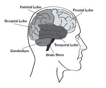 |
| Cranial Nerves 12 pairs of nerves come directly out of the brain (cranium) instead of the spinal cord, so they're called cranial nerves. They each have specific functions: Cranial Nerve I (Olfactory) - transmits sense of smell from the nasal cavity to the brain [ol-fak´tŏ-re] Cranial Nerve II (Optic) - visual signals from the retina to the brain Cranial Nerve III (Oculomotor) - controls eyeball movement, pupil dilation and upper eyelid Cranial Nerve IV (Trochlear) - controls eyeball movement [trok´le-ar] Cranial Nerve V (Trigeminal) - sensation of the face and muscles of chewing (mastication) [tri-jem´ĭ-nal] Cranial Nerve VI (Abducens) - eyeball movement (specifically, the lateral rectus muscle) [ab-du´senz] Cranial Nerve VII (Facial) - facial expression, ear ossicles, front part of tongue sense of taste Cranial Nerve VIII (Acoustic) - hearing and sense of balance Cranial Nerve IX (Glossopharyngeal) - sense of taste and sensation of back of throat [glos″o-fah-rin´je-al] Cranial Nerve X (Vagus) - throat muscles and parasympathetic nerves to the chest and abdomen organs (va´gus) Cranial Nerve XI (Accessory) - sternocleidomastoid and trapezius muscle Cranial Nerve XII (Hypoglossal) - muscles of the tongue [hi″po-glos´al] |
 |
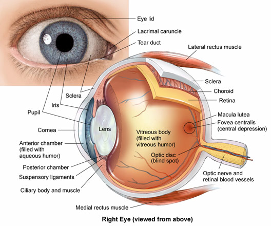 |
SIGHT: Light enters the eye through the clear lens. The pupil widens or narrows to control how much light can enter. The inner surface of the eye, called the retina, contains the specialized sensory cells called rods (light/dark) and cones (color) that send the visual signal to the occipital lobe through the optic nerve (Cranial Nerve II). Movement of the eyeball is controlled by the extraocular muscles. |
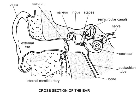 |
HEARING: Sound waves travel through the ear canal and strike the tympanic membrane (ear drum) which vibrates, moving the 3 tiny bones, or ossicles, called the malleus, incus, and stapes. The vibrating ear drum is attached to the malleus which moves the incus which moves the stapes [stā′pēz]. When the stapes moves it stimulates the cochlea [kok′lē·ə], which senses the sound. The sound signal is sent to the temporal lobes through the Acoustic Nerve (Cranial Nerve VIII). The sense of balance is regulated by the 3 semicircular canals in the inner ear. |
| Multiple Sclerosis (MS) - the myelin sheath that covers [delicious] brain neurons is inflamed, resulting in all kinds of sensory and motor abnormalities such as numbness, tingling, pain, fatigue, weakness of arms or legs, slurred speech, double vision, etc. With almost any neurologic symptom a patient complains of, the diagnosis of MS could be considered. On the MRI x-ray it will appear as abnormal white areas in the [delicious] brain. |  |
| Subdural hematoma - when bleeding develops inside the skull (usually from an head injury), blood can collect underneath the dura mater layer of the meninges, i.e. "sub" (under) "dural" (dura): it develops in the space between the [delicious] brain and the dura mater. A collection of blood anywhere is called a hematoma and a bruise is a type of hematoma. It often appears in a football-shape area on CT scan x-ray. A similar kind of bleeding is called a subarachnoid hemorrhage. It can be caused by rupture of a dilated blood vessel in the brain called an aneurysm. Obviously, anytime there's bleeding here it can be bad. |  |