The Development of Technology and Its Influence on Nuclear Medicine
by Tess Hughes


1950-59
|
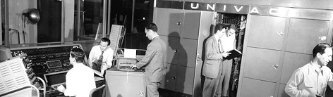
In 1951, Eckert and Mauchly developed the world's first
general-purpose electronic digital computer, the UNIVAC (UNIVersal Automatic Computer).
Instead of flashing lights, results could be printed out on paper.
It was the first computer to use magnetic tape rather than International Business Machines
(IBM's) slower punch card technology. UNIVAC became a household name in
1952 by predicting Eisenhower's presidential election and so
IBM retaliated. They offered 60% discount for educational uses and quickly dominated the
university market.4
The strong relationship between education and computing was forged.
|

Figure 35
The UNIVAC
|
|
Also in 1951, the beginnings of Nuclear Medicine arose with the rectilinear scanner.
Invented by Benedict Cassen, it produced an image on paper copying the scanning motion
of the detector but was prone to jamming.

Figure 47
George Mueller
|
|
In 1952
George E.Mueller developed a more reliable system, by arcing each amplified pulse through
layered paper to burn a black spot thus forming an image.6
In 1953 Hal Anger developed the first recognised
'gamma camera' in the AEC Donner Lab at Berkley, USA. It produced images as an
array by using a single, thallium impregnated sodium iodide (NaI(Tl)) screen and
a sheet of x-ray film rather than in the linear fashion produced by the rectilinear
scanner. It was however, very time-consuming, taking up to an hour for each image
with therapeutic doses of radioactivity.4
|


|
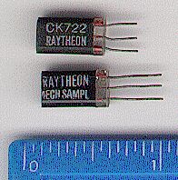
The Raytheon CK722 Junction Transistor, shown at the top of this old photograph,
was the first mass-produced transistor and was manufactured in
1953.1
In 1954, Bell Laboratories built the first 'Second Generation Computer'
containing transistors. The more complex the circuits became, however, the more complicated and
numerous were the soldered connections between transistors, and the likelihood of faulty
wiring increased.37
|

Figure 5
The Raytheon CK722 transistor1
|
|
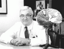 Figure 68
David Kuhl
|
|
It was not until 1956 that David Kuhl developed photographic output for the
rectilinear scanner. This utilised a glow-lamp and x-ray film and accentuated
differences in intensity of detector signal, therefore producing images of better
contrast. These advances allowed dynamic imaging of blood flow and function,
rather than purely static information.6
|
|
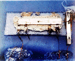
In 1958, Jack St. Clair Kilby of Texas Instruments, manufactured the first integrated
circuit or chip. This minimised the faulty wiring problem of transistor circuits by
connecting the tiny transistors during chip manufacture. The diminished distance
between the transistors dramatically increased the speed of computers as well. The
only connections needed were between the chips and other electronic components.38
|

Figure 79
First integrated circuit
|
|

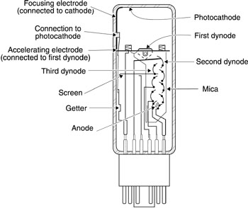
Figure 811
Typical photmultiplier tube assembly
|
|
Also in 1958, Anger replaced the film-screen combination with a single, large
field-of-view NaI(Tl) crystal and photomultiplier tube (PMT) assembly. This resulted
in far greater detection efficiency. The PMT array, combined with electronic position
logic circuitry, determined the location of each scintillation event as it occurred
in the crystal.10
|
|
In addition to this, the energy of the scintillations could be analysed and if
outside a predetermined energy range, could be excluded from further calculation
and cathode ray tube (CRT) display. The CRT display showed a pattern of light
that corresponded to the scintillations occurring in the detector crystal. These
brighter flashes of light were more easily recorded photographically. This improved
resolution, and reduced the time of acquisition and the radiation dose required.
The images were recorded on Polaroid® Film or on x-ray film, which was attached to the
camera housing.10
|
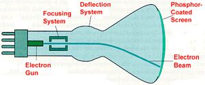
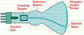 Figure 912
The cathode ray tube
|
|
The gamma camera made its debut at a Los Angeles Nuclear Medicine Society meeting
in 1958. Acceptance was slow, because manufacturers were developing and improving
the rectilinear scanner. Initially using a pinhole collimator, Anger developed the
multi-hole collimator. This resulted in even better image resolution when coupled
with larger sodium iodide crystals and more PMT's of better quality.13
See Figure 11.
|