

 Serial sections up through clusters of P19 neurons.
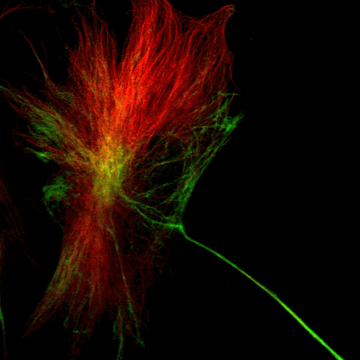
Protein 4.1
Tubulin
Colocalisation
 Two sections of ND cells (basal and lateral). In the lateral section a number of cells can be seen in mitosis, characterised by a change in the shape and pattern of colour (rounder with red centre and green surround).
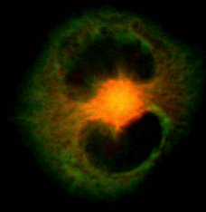 A serial section up through a single ND cell. The kidney-shaped nucleus is visible in black surrounding the microtubule-organizing centre (MTOC) in yellow.
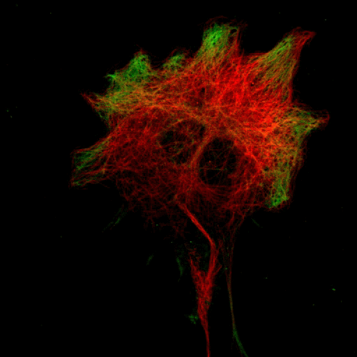
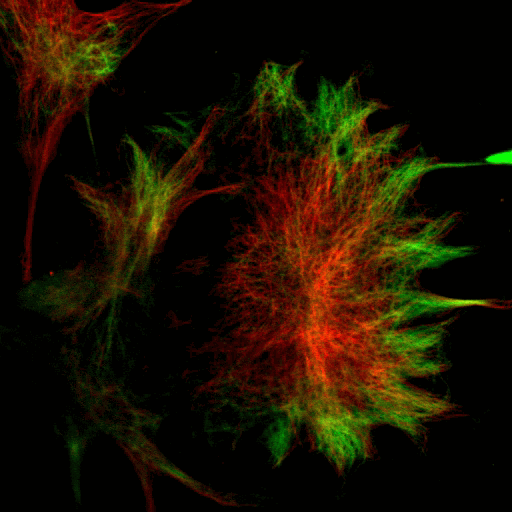
 Protein 4.1


Protein 4.1
Tubulin
Colocalisation
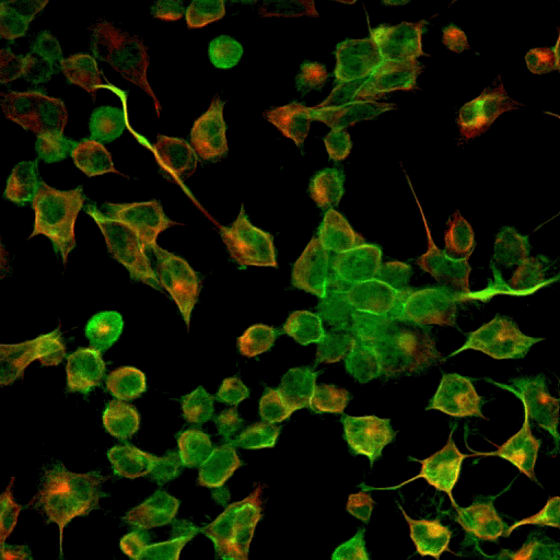
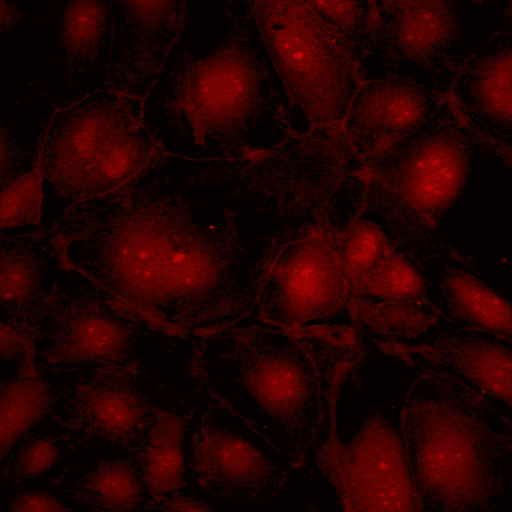 Untidy cell-cell contacts where membrane sheets overlap are highlighted by anti-P4.1.
Protein 4.1
Actin
Colocalisation
