If you have diabetes mellitus, you probably know that your body can't use or store sugar properly. When your blood sugar gets too high, it can damage the blood vessels in your eyes. This damage may lead to diabetic retinopathy. (Your retina is the nerve layer that lines the inside of your eye and converts light into nerve signals that your brain can interpret.)
.
Types Of Diabetic Retinopathy
When blood vessels in the retina are damaged, they can leak fluid or bleed. This causes the retina to swell and form deposits called exudates.
This is an early form of diabetic retinopathy called nonproliferative or background retinopathy. You may not notice any change in your vision when you develop this early form of the disease, but it can lead to other more serious forms of retinopathy that affect your vision.
When fluid collects in the macula (the part of the retina that allows us to see fine details), reading and other close work may become difficult, This is called macular edema.
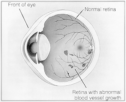
In proliferative retinopathy, new fragile blood vessels grow on the surface of the retina.
These new blood vessels are called neovascularization, and can lead to. serious vision problems, because the new vessels can break and bleed. into the vitreous. (The vitreous is the clear, jelly-like substance. that fills the center of the eye.)
When the vitreous becomes clouded with blood, light is prevented from passing through the eye to the retina. This can blur or distort vision.
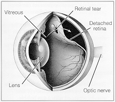
The new blood vessels can also cause scar tissue to develop, which can pull the retina away from the back of the eye.
This is known as retinal detachment, and can lead to blindness if untreated. In addition, abnormal blood vessels can grow on the iris (the colored part in the front of your eye), which can lead to glaucoma.
What is the best defense against diabetic retinopathy?
Early treatment of retinopathy often improves the potential for saving sight. Every person with diabetes should receive regular care from a doctor and closely follow the prescribed treatment plan.
It is also very important to regularly visit an eye care professional even before any visual symptoms appear. An eye examination through dilated pupils by an opthomologist should be repeated at least once a year for both adults and children who have diabetes.
Parts Of The Eye
Cornea - The clear front window of the eye. The cornea transmits and focuses light
into the eye.
Iris - The colored part of the eye. The iris helps regulate the amount of light that
enters the eye.
Pupil - The dark center in the middle of the iris. The pupil determines how much
light is let into the eye. It changes sizes to accommodate for the amount of light that
is available.
Lens - The transparent structure inside the eye that focuses light rays onto the retina.
Retina - The nerve layer that lines the back of the eye. The retina senses light and
creates impulses that are sent through the optic nerve to the brain.
Macula - A small area in the retina that contains special light-sensitive cells. The
macula allows us to see fine details clearly.
Optic Nerve - The nerve that connects the eye to the brain. The optic nerve carries
the impulses formed by the retina to the brain, which interprets them as images.
Vitreous - The clear, jelly-like substance that fills the middle of the eye.
Visit The Links Below For Some Informative Information On Eyes.
We subscribe to the HONcode principles. Verify here.
This site supports the Health on the Net Code of Conduct to improve the quality of the Medical Internet.
NOTICE: The medical information and the links on this website is maintained voluntarily for the benefit of those with an interest in diabetes. I am not a medical professional. The information here reflects my personal experiences. Where appropriate, consult your physician before changing your diabetic treatment plan.