
HEART
DISEASES
Disorders of the heart kill more
people in developed nations than any other disease. They can arise from
congenital defects, infection, narrowing of the coronary arteries, high blood
pressure or disturbances of heart rhythm.

Congenital heart defects include
persistence of fetal connections between the arterial and venous circulations
such as the ductus arteriosus a vessel normally connecting the pulmonary artery
and the aorta only until birth. Other important developmental anomalies involve
the partition separating the four cardiac cavities and the large vessels
issuing from them. In newborn “blue babies”,
the pulmonary artery is narrowed and the ventricles are connected by an
abnormal opening; in this cyanotic condition, the skin has a bluish tinge
because the blood receives an insufficient amount of oxygen. Formerly the
expectation of life for such infants was extremely limited; with the advent of
early diagnosis and improved techniques of hypothermia, surgery is often
possible in the first week of life and the outlook for these infants greatly
improved.
Rheumatic heart disease was formerly
one of the most serious forms of heart disease of childhood and adolescence,
involving damage to the entire heart and its membranes. It usually followed
attacks of rheumatic fever. Widespread use of antibiotics effective against the
streptococcal bacterium that causes rheumatic fever has greatly reduced the
incidence of this condition.
Myocarditis is inflammation or
degeneration of the heart muscle. Although it is  often caused by various diseases
such as syphilis, toxic goitre, endocarditis, or hypertension, myocarditis may
appear as a primary disease in adults or as a degenerative disease of old age.
It may be associated with dilation or with hypertrophy.
often caused by various diseases
such as syphilis, toxic goitre, endocarditis, or hypertension, myocarditis may
appear as a primary disease in adults or as a degenerative disease of old age.
It may be associated with dilation or with hypertrophy.
The major form of heart disease in
Western countries is atherosclerosis. In this condition fatty deposits called
plaque, composed of cholesterol and fats, build up on the inner wall of the
coronary arteries. Gradual narrowing of the arteries throughout life restricts
the blood flow to the heart muscles. Symptoms of this restricted blood flow can
include shortness of breath, especially during exercise, and a tightening pain
in the chest called angina pectoris. The plaque may become  large enough to completely obstruct
the coronary artery, causing a sudden decrease in oxygen supply to the heart.
Obstruction, also called occlusion, can occur when part of the plaque breaks
away and lodges farther along in the artery, a process called thrombosis. These
events are the major causes of heart attack, or myocardial infarction, which is
often fatal. Persons who survive a heart attack must undergo extensive
rehabilitation; there is always the risk of a recurrence.
large enough to completely obstruct
the coronary artery, causing a sudden decrease in oxygen supply to the heart.
Obstruction, also called occlusion, can occur when part of the plaque breaks
away and lodges farther along in the artery, a process called thrombosis. These
events are the major causes of heart attack, or myocardial infarction, which is
often fatal. Persons who survive a heart attack must undergo extensive
rehabilitation; there is always the risk of a recurrence.
Development of fatty plaque is due
partly to excessive intake of cholesterol and animal fats in the diet. A
sedentary life-style is thought to promote atherosclerosis, and evidence
suggests that physical exercise may help prevent heart disease. A striving,
perfectionist temperament referred to as Type A personality
has also been associated with  increased risk of heart attacks as
has cigarette smoking. The occurrence of the heart attack itself is much more
likely in persons who have high blood pressure. The actual event precipitating
the attack may involve products secreted by platelets in the blood. This has
led to clinical studies testing whether persons who have had a heart attack
will be protected from a second infarction if they take drugs that block the
action of platelets.
increased risk of heart attacks as
has cigarette smoking. The occurrence of the heart attack itself is much more
likely in persons who have high blood pressure. The actual event precipitating
the attack may involve products secreted by platelets in the blood. This has
led to clinical studies testing whether persons who have had a heart attack
will be protected from a second infarction if they take drugs that block the
action of platelets.
Many persons having severe angina
because of atherosclerotic disease can be treated with drugs, such as beta
blockers (for example, propranolol) and nitrates, which reduce the load on the
heart. Those who do not obtain relief with pharmacologic means can often be
treated by a form of surgery called coronary bypass. In this procedure, which
became established in the 1970’s, a section of vein from the leg is sewn into
the blocked coronary artery to form a bridge around the atherosclerotic region.
In most recipients the operation relieves the pain of angina and in many
persons it prevents a fatal heart attack.
A second surgical procedure that was
developed during the 1970’s to treat atherosclerotic heart disease is balloon
catheterization, technically called percutaneous transluminal coronary
angioplasty. In this operation a wire with a balloon on the tip is inserted
into an artery in the leg and threaded through the aorta into the coronary
artery. When the balloon reaches the atherosclerotic area, it is inflated. The
plaque is compressed and normal blood flow is reestablished. It is estimated
that about one in six coronary bypass operations can be replaced by this less
dangerous procedure.
During the 1970’s and early 1980’s
it became apparent that a dramatic drop was occurring in mortality from
atherosclerotic heart disease in several developed countries. Although no
definitive explanation for this decline has been given, public health officials
have attributed it to widespread detection and treatment of high blood pressure
and a decrease in the amount of animal fat in the average Western diet.
Some persons who die of apparent
heart attack exhibit no evidence of severe athero-sclerosis. Research has shown
that a decrease in blood flow to the heart can also be from the spontaneous
contraction of an apparently healthy coronary artery (vasospasm) which may
contribute to some heart attacks brought on by atherosclerosis.
The immediate cause of death in many
heart attacks, whether atherosclerosis is present or not, is ventricular
fibrillation—cardiac arrest. This is a rapid ineffective beating of the
ventricles. Normal heart rhythm can often be restored by a massive electric
shock to the chest, a finding that has led to emergency rescue teams in many
cities being trained in this technique.
Minor variations in the heart rhythm
usually have little pathological significance. The heart rate responds to the
demands of the body over such a wide range that variations are generally within
normal limits. Severe defects however in the sinoatrial node or in the fibers
that transmit impulses to the heart muscle can cause dizziness, faintness and
eventually death. The most serious of these conditions is called complete heart
block. It can be corrected by insertion of an artificial pacemaker, a device
that gives timed electric shocks to make the heart muscle contract in a regular
pattern. Most other arrhythmias are not dangerous except in persons with
underlying heart disease. In these patients, especially those who have already
had a heart attack, arrhythmias are treated with propranolol, lidocaine and
disopyramide.
Often found among older persons is
pulmonary heart disease, which is usually the result of a lung ailment such as
emphysema, or a disease affecting circulation to the lungs, such as
arteriosclerosis of the pulmonary artery. Another condition found in older
persons is congestive heart failure, in which the ventricles pump far less
efficiently. The muscular walls of the ventricles enlarge with the effort to
propel more of the blood into the circulation, giving rise to the large, floppy
hearts characteristic of this syndrome. Persons with this ailment have a
reduced capacity for exercise. Their condition can often be improved with one
of the derivatives of digitalis, which increases the pumping efficiency of the
heart.
Diagnosis
The electrocardiograph is an
instrument for recording the electrical currents produced by the heart muscle
during various phases of contraction and an important diagnostic tool. The
efficiency of the heart as a pump may be measured accurately by the use of
cardiac catheterization. In this technique a tube is introduced, through a vein
or an artery or both, into the right, left, or both heart cavities, the
pulmonary artery, and the aorta. This process permits determination of the rate
of blood flow and recording of blood pressure in intracardiac and large
vessels. This technique makes it possible to detect abnormal communications
between right and left heart cavities. In another diagnostic technique called  angiocardiography, or cinefluoroscopy,
photographic recordings are obtained of the heart cavities and of the pathways
and contours of the pulmonary vessels and the aorta with its branches; the
technique involves injecting a substance opaque to X-rays into a vein. Even
more accurate delineation of areas of reduced blood flow in the heart is
provided by a new technique that visualizes the flow of a radioactive isotope
of the element thallium into heart muscle. A computerized camera records the
extent of thallium penetration during the systole-diastole cycle of the heart,
precisely showing small areas of tissue damage. Yet another technique that is
now being used is ultrasound—ultrasonic imaging.
angiocardiography, or cinefluoroscopy,
photographic recordings are obtained of the heart cavities and of the pathways
and contours of the pulmonary vessels and the aorta with its branches; the
technique involves injecting a substance opaque to X-rays into a vein. Even
more accurate delineation of areas of reduced blood flow in the heart is
provided by a new technique that visualizes the flow of a radioactive isotope
of the element thallium into heart muscle. A computerized camera records the
extent of thallium penetration during the systole-diastole cycle of the heart,
precisely showing small areas of tissue damage. Yet another technique that is
now being used is ultrasound—ultrasonic imaging.
Heart Transplants
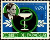 In 1967 a human heart from one
person was transplanted into the body of another by the South African surgeon Christiaan Barnard. Many surgeons have
since adopted the procedure. The major problem at first was the body's natural
tendency to reject tissues from another individual. By the
In 1967 a human heart from one
person was transplanted into the body of another by the South African surgeon Christiaan Barnard. Many surgeons have
since adopted the procedure. The major problem at first was the body's natural
tendency to reject tissues from another individual. By the  early 1980’s however due to the use
of immunosuppressive drugs, particularly cyclo-porine, many more cardiac
transplant recipients were living beyond one year. By the 1990’s the operation
had become more commonplace in developed nations such as the
early 1980’s however due to the use
of immunosuppressive drugs, particularly cyclo-porine, many more cardiac
transplant recipients were living beyond one year. By the 1990’s the operation
had become more commonplace in developed nations such as the
Christiaan Neethling Barnard 1922 -
Is South African surgeon, who
performed the first human heart-transplant operation. Barnard
was born in Beaufort West and received an MD degree from the
![]()
