|

|
|
|
|
| |
|
This page is an accumulation of information from numerous sources(Internet, research, newspapers, etc.). The list is for information purposes only and should not replace proper diagnosis from a physician.
|
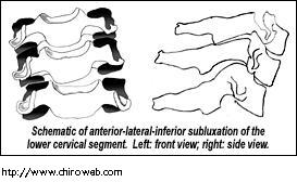
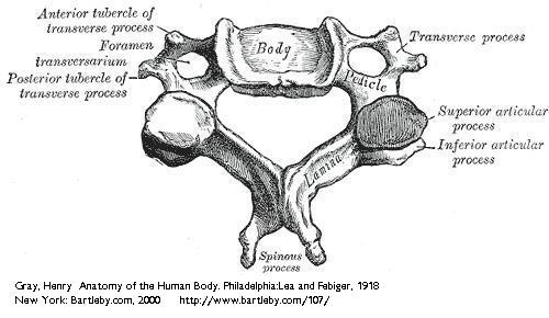
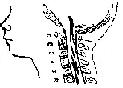
Atlanto-axial subluxation:
Instability of the first and second vertebrae in the neck. It takes its name from the proper name of the first cervical vertebrae (the atlas) and the second vertebrae (the axis). The cause may include abnormalities of the ligaments, or bony abnormalities of the cervical vertebrae or both. Indications for surgery include neurologic abnormality with instability, intractable neck and head pain, vertebral artery compromise, and cord compression on MRI (even without symptoms). The most common surgical procedure for anterior C1-C2 subluxation is posterior fusion by internal fixation of C1-C2.
|
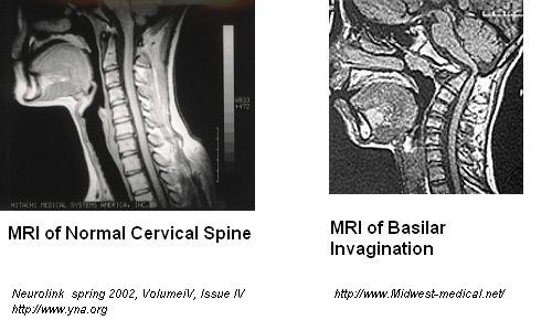
Basilar Invagination:
Is diagnosed when the superior part of the odontoid (part of the C2 vertebrae) migrates upward. It is uncommon but somewhat dangerous. It occurs both congenitally and in persons with bone diseases such as rheumatoid arthritis. It may lead to static or dynamic stenosis of the foramen magnum and compression of the medulla oblongata (lower brainstem). A closely related condition, the Chiari malformation, mainly occurs congenitally. Neurosurgery is recommended when neurologic symptoms and signs are present and cord compression is confirmed by MRI. When these features are absent a conservative approach may be pursued such as a collar, NSAID's, and simple neck traction.

|
Chiari Malformation:
A brain malformation characterized by a small or misshapen posterior fossa (compartment in the back of the skull), and a protrusion of the structures in the back of the brain(cerebellar tonsils) into the spinal canal. Chiari Malformations were first described in the 1890's by a German pathologist, professor Chiari. He assigned a grade to the malformations: beginning with type one the mildest form through type four the most severe. A colleague, Dr. Arnold, added to the type two description and hence the name "Arnold- Chiari Malformation".
|
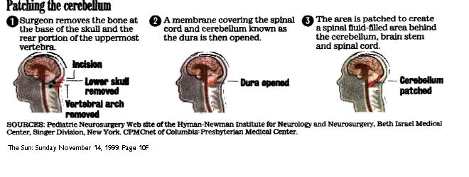
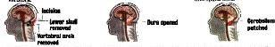
Decompression:
A surgical procedure in which the surgeon removes the bone at the base of the skull(craniotomy) and the rear portion of the uppermost vertebra(Laminectomy). Some surgeons then open up the thin membrane that covers the brain and cerebellum called the Dura. Once this area is opened, a patch must be made to close it (Dura Graft). The patch can be made from bovine, cadaver, synthetic, periosteum(scalp), or pericardium. Techniques and procedures vary depending on the surgeon.
|
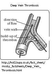
Deep Vein Thrombosis:
Refers to the formation of a Thrombus(blood clot) within a deep vein. The blood clot can either partially or completely block the flow of blood in the vein. Symptoms include, tenderness and redness in the effected area, pain and swelling in areas drained by the vein where the clot is located, fever, rapid heart beat, sudden unexplained cough, joint pain and soreness. Treatment includes applying warm moist heat to relieve pain. Medications are often prescribed called anti-coagulants or anti-platelet agents. These drugs help prevent more clots from forming(Warfarin, Heparin etc.)

|
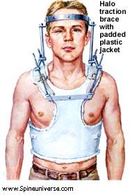
Halo Traction:
In Spinal Traction, parts of the spinal column are pulled in opposite directions in order to stabilize or change the position of damaged aspects of the spine. The force is usually applied to the skull through a series of weights or a fixation device and requires that the patient is either kept in bed or placed into a Halo vest. Traction is used for several spinal problems including mobilization of soft tissues or joints, decompression of pinched nerve roots, and reduction of herniated intervertebral disks. Most importantly it is used for the management of spinal instability. The Halo Brace is often used as the initial treatment of odontoid fractures. A Halo metal ring is secured to the skull with pins and to two metal rods attached to a well fitted plastic jacket. The Halo obtains complete fixation and arrests almost all movement of the cervical spine.

|
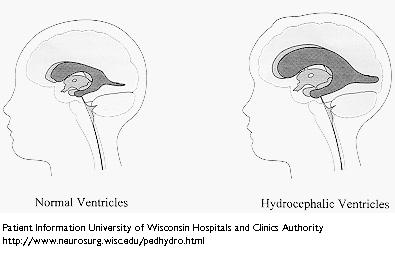
Hydrocephalus:
An abnormal accumulation of fluid, cerebrospinal fluid or CSF, within the cavities of the brain called ventricles. CSF surrounds the brain and spinal cord and acts as a protective cushion against injury. Hydrocephalus occurs when there is an imbalance between the amount of CSF produced and the rate that it is absorbed by the blood stream. As CSF builds up, it causes the ventricles to enlarge and the pressure inside the head to increase. Hydrocephalus may be congenital(present at birth), or acquired as a result of Spina Bifida, Meningitis, Head Trauma, Tumors, Cysts, etc. There is no known way to prevent or cure Hydrocephalus. Treatment involves the surgical insertion of a shunt.

|
Laminectomy:
A surgical procedure designed to relieve pressure on the nerves in either the back or the neck. For a cervical Laminectomy, an incision is made either in the back of the neck (posterior cervical) or in the front side of neck (anterior cervical) depending on the location of the problem. The bone that curves around and covers the spinal cord (lamina) is then removed and the tissue that is causing pressure on the nerves or spinal cord is also removed. The hole through which the nerve passes may also be enlarged to prevent further pressure on the nerve.

|
Minerva Brace:
A Brace that immobilizes the spine from C1 to T1. It controls flexion- extension and rotation of the spine.
In Roman mythology, Minerva was the goddess of wisdom, commerce, crafts, and the inventor of music. Ovid called her the "goddess of a thousand words." Romans celebrated her worship from March 19- 23 during Quinquatrus, the artisans holiday. Minerva's Greek counterpart is Athena. Both were said to have sprung fully grown and fully armored from the Head of Jupiter(Zeus).


|
Orthostatic Hypotension:
The sudden drop in Blood Pressure that occurs when a person assumes a standing position. There are several causes including not taking in enough fluids or salt, some medications, and some hormonal problems. However, much of the time the problem is due to the nerves not telling the vessels to close down when you stand up. Symptoms which generally occur after sudden standing include dizziness, lightheadedness, blurred vision, and syncope(temporary loss of consciousness). Medications are available- Fludrocortisone (Florinef) improves vessel response at low dose and causes fluid retention in high doses. Midodrine (Proamatine) directly causes the vessels to close down by acting just like the chemical that the nerves would release if they were functioning properly. Beta Blockers (Inderal) prevent the veins from opening excessively and may also trigger an increased standing Blood pressure through unknown mechanisms.
|
Occupational Therapy:
The therapeutic use of work, self care, and play activities to increase independent function, enhance development, and prevent disability. It may include adaptation of task or environment to achieve maximum independence and enhance quality of life.
|
Pneumothroax:
Collapsed lung. Occurs when air enters the space between the two layers of the pleura(membrane that surrounds the lungs). The air separates the two layers and causes part or all of the lung to collapse and lose air that is normally inside the lung.
|
Sepsis:
A serious infection that is caused by bacteria that has entered a wound or body tissue that leads to the formulation of pus or to the spread of bacteria in the blood. Symptoms include: fever, hyperventilation, chills, shaking, warm skin, skin rash, rapid heart beat(tachycardia), confusion, delirium, decreased urine output.
|
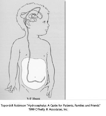
Shunt:
A device that is used to divert the buildup of cerebrospinal fluid(CSF) to another area of the body where it can be reabsorbed. Subarrachnoid- The area between the Pia and arrachnoid matter known as the subarrachnoid space, where cerebral spinal fluid flows over the surface of the brain and spinal cord. VP (Ventriculoperitoneal shunt)- Diverts CSF away from the ventricular system of the brain and drains into the peritoneal cavity. The peritoneum lines the abdominal cavity, which is where the digestive organs are located(i.e., stomach and intestines).The CSF is then absorbed into the bloodstream by blood vessels in the wall of the abdomen.

|
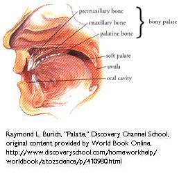
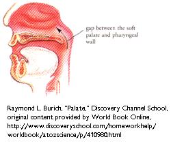
Soft Palate:
The Palate (roof of the mouth) is a wall partitioning the nasal and oral cavities. This partition is made up of two parts, the Hard and Soft Palate. The Hard Palate forms the front portion and is made up of two bony plates. The Soft Palate forms the rear portion of the Palate wall. The Soft Palate consists of muscles and typically elevates during speech and swallowing to separate the nose and mouth.

|
Syringobulbia:
Syringobulbia is a neurological disorder characterized by a fluid-filled cavity (syrinx) within the spinal cord that extends to involve the brain stem. It usually occurs as a slitlike gap within the lower brainstem that may affect the lower cranial nerves including sensory and motor nerve pathways by disruption or compression.
|
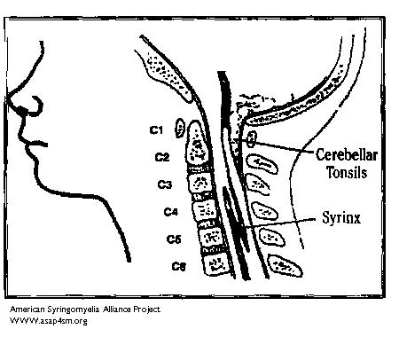
Syringomyelia:
A chronic spinal disorder in which cerebrospinal fluid enters the spinal cord, forming a cavity known as a syrinx. Doctors sometimes use other words such as cyst, hydromyelia, or syringohydromyelia. In most cases syringomyelia(SM) is related to a congenital malformation (Chiari). However, not all patients with Chiari Malformation will develop a syrinx. SM can also occur as a complication of spinal trauma, meningitis, tumor, or arachnoiditis. In these cases a syrinx develops in the section of the spinal cord damaged by these conditions. The symptoms of SM are numerous and a person may have various combinations of symptoms. The common symptoms include: loss of sensitivity, especially to hot and cold, muscle weakness and spasticity, motor impairement, loss of bladder and bowel control. Sensory problems such as numbness, tingling, burning and sweating problems may occur. The majority of patients suffer from headaches and chronic pain.
|
Syrinx:
A fluid filled cavity within the spinal cord. A syrinx may expand and elongate in time, destroying the center of the spinal cord. As the nerve fibers inside the spinal cord are damaged, a wide variety of symptoms can occur, depending on the size and location of the syrinx.
|
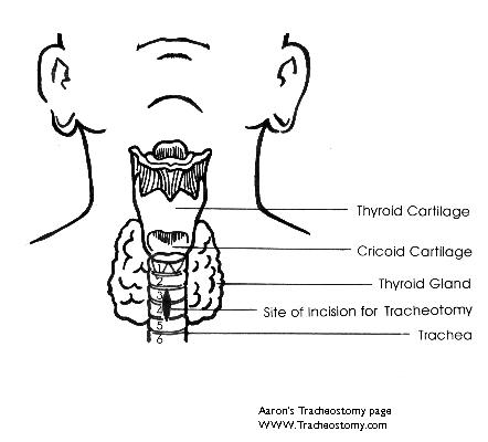
Tracheostomy:
A surgical procedure in which an incision is made into the trachea(windpipe) that forms a temporary or permanent opening. A tube is inserted through the opening to allow the passage of air and removal of secretions.

|
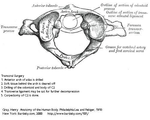
Transoral surgery:
A surgical procedure that is done through the mouth. The transoral approach is used to treat Basilar Invagination, Rheumatoid Arthritis with Atlanto-axial subluxation, Odontoid fractures with Atlanto-axial subluxation, and other disorders of the upper cervical spine. When used for Basilar Invagination, the odontoid (upper portion of the second cervical vertebrae) is removed. A cervical fusion is usually performed after to provide stability.
|
Velopharyngeal Insufficiency:
The improper closing of the velopharyngeal sphincter (soft palate muscle) during speech. It is characterized by an acute nasal quality of the voice. The primary symptom is the speech impediment. Some people develop a change in their speaking pattern or a series of facial grimaces to try to overcome the difficulty. If the condition is acute, regurgitation through the nose may occur. Velopharyngeal insufficiency is treated with a combination of surgery and speech therapy. The combination of surgery to correct the insufficiency, and speech therapy to retrain the voice, successfully alleviates the condition.

|
Surgery 1986:
Suboccipital craniectomy with duraplasty, Laminectomy C1-C2-C3 removal of the arc,laminae, and spinous processes, Decompression, insertion of right Ventricular Peritoneal shunt.
|
Surgery 1999:
Transoral Transpalatopharyngeal resection of anterior atlas arch, clivus odontoid process and ventral cervicomedullary decompression. Posterior fossa exploration, intradural lysis of adhesions with resection of left cerebral tonsil, cervical fascia duraplasty. Dorsal occiput-C2-C3-C4 fusion with custom contoured threaded titanium loop with titanium cables and autologous rib graft.
|
Surgery 2000:Posterior fossa exploration, Subarrachnoid shunt, Periumbilical Fat graft and fat injection into the nasopharynx.
|
Surgery 2001:Emergency tracheostomy,transpalatalpharyngeal resection of the C2 body with medullary decompression.
|
|