THANK YOU FOR STOPING BY, THIS PAGE WILL BE ABOUT THE DISEASE THAT KAIN SUFFERS FROM, HYDRONEPHROSIS. I AM PLANNING ON GIVING AS MUCH MEDICAL INFORMATION ABOUT THIS DISEASE AS I CAN FIND. BEFORE MY SON WAS BORN, I COULDN'T EVEN PRONOUNCE THIS WORD, NOW I AM LEARNING AS MUCH AS I CAN, AND WANT TO EDUCATE OTHERS ABOUT THIS.
Can you tell me more about hydronephrosis?
Hydronephrosis is being detected before birth with greatly increasing frequency. In the recent past, hydronephrosis (an enlargement of the kidneys caused by some degree of blockage to normal urine flow) was uncommon and was usually treated surgically. Most of the cases of hydronephrosis were detected due to either decreased amniotic fluid or decreased urine output after birth. If left untreated hydronephrosis would cause progressive kidney damage.
Today the situation is entirely different. With an explosion of the number and quality of prenatal ultrasounds, a huge number of children with some degree of hydronephrosis have been discovered. In some studies as many as 2% of children (mostly boys) have been diagnosed with prenatal hydronephrosis.
This sudden increase in the number of cases of hydronephrosis has led to a flurry of controversy and confusion. What is the real significance of this asymptomatic hydronephrosis? How do we know which newborns with hydronephrosis will get worse and which will stabilize or improve? When should we operate on these children, and when should we choose to observe them? Which children have hydronephrosis caused by a urine obstruction early in development that has already corrected itself? In which children is there still an obstruction to urine flow? These dilemmas are currently being sorted out.
We do know that about half of the cases of prenatal hydronephrosis will resolve spontaneously before the child is even born. In one study of children in which the prenatal hydronephrosis persisted after birth, continued follow-up showed that 93% of the children had complete resolution of their hydronephrosis over time -- with no loss of kidney function. Only 7% of those who still had the hydronephrosis after birth went on to have progression of the hydronephrosis and eventually required surgery. Those who did require surgery did not have any permanent loss of kidney function.
The management of children with hydronephrosis is still evolving. Most agree that a repeat ultrasound shortly after birth should be done to assess the progression of the condition. If the hydronephrosis is stable or improving and there is no current obstruction identified, most doctors recommend managing the child conservatively with close follow-up and prophylactic antibiotics to prevent kidney infections. If at any point an ongoing obstruction is found or if the hydronephrosis is worsening, surgery may be needed. Thankfully this surgery is safe and effective.
So for your child, one of two situations pertains:
Probably your baby has a benign condition that a few years ago never would have been noticed at all, and which will resolve spontaneously. This condition will cause no bigger problem than your short-term worry and concern.
Possibly your child has a hydronephrosis that results from an ongoing obstruction. In this case finding it early will help to get the needed treatment at the optimum time.
The above information is taken from Dr. Green's House Calls, Pediatric Wisdom for the Information Age
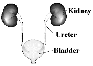
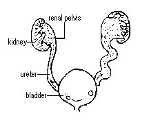
So, what does hydronephrosis look like? Here is a picture of what the "normal" kidneys look like..
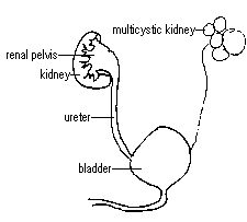
Now, take a look a kidney suffering form hydronephrosis....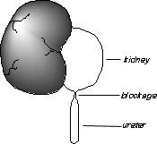
In extremely severe cases of hydronephrosis, damage to normal kidney function may occur. In addition to affecting the child's kidney function, this condition may also cause infections, pain, and bleeding. These effects may take months or even years to occur or may never occur.
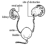
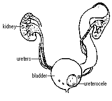
Take a look at the below pictures, showing what severe hydronephrosis looks like...
The first picture shows a normal kidney, the second shows a kidney suffering from "Mid" Hydronephrosis, the third shows a kidney suffering from "Moderate" Hydronephrosis, and the fourth shows a kidney suffering from "Severe" Hydronephrosis.

The kidneys, located in the back of the abdomen just below the ribs, are important for many bodily functions. One of the kidneys' major functions is to filter blood and remove waste products that are passed out of the body through the urine. The kidneys are connected to thin tubes called ureters, which carry the urine to the bladder. When the kidneys are forming before a child is born, sometimes conditions develop that change the way one or both of the kidneys look or function. One such condition is hydronephrosis.
Hydronephrosis is a "stretching" or dilation of the inside or collecting part of the kidney. It often results from a blockage in the ureter where it joins the kidney that prevents urine from draining into the bladder. Urine is trapped in the kidney and causes the kidney to stretch. Hydronephrosis may also be due to abnormal backwash or "reflux" of urine from the bladder. In infants the amount of hydronephrosis may appear greater than the actual degree of blockage because of the "stretchiness" of the young tissues.
Neonatal hydronephrosis is often detected on an ultrasound test during pregnancy (prenatal ultrasound). Hydronephrosis has never been linked to anything the parents have done during pregnancy and is usually not passed from parent to child or found in more than one sibling in a family. The blockage that produces hydronephrosis is usually the result of a narrowing at the top of the ureter near the kidney that probably developed before the fourth month of pregnancy.
The severity of hydronephrosis depends on the extent of the blockage and the amount of stretching of the kidney. Hydronephrosis may range from mild to severe. Children with mild hydronephrosis usually do not have symptoms, the kidneys are minimally affected, and the hydronephrosis may disappear in the first year of life.
In patients observed with moderate hydronephrosis, kidney function usually does not decrease, kidney growth remains normal, and the degree of hydronephrosis usually does not worsen. Research has shown that careful observation during the first one to two years of life suggests that the kidney compensates for the hydronephrosis to maintain normal function and that some children even get better on their own.
In extremely severe cases of hydronephrosis, damage to normal kidney function may occur. In addition to affecting the child's kidney function, this condition may also cause infections, pain, and bleeding. These effects may take months or even years to occur or may never occur.
To determine the amount of hydronephrosis present, "nuclear medicine" and radiologic tests to measure kidney function and structure may be recommended by the physician.
In children with mild hydronephrosis, observational therapy has been shown to be safe and has become the accepted method of treatment. However, in children with moderate to severe hydronephrosis, the answer is not as clear.
The surgery to correct hydronephrosis is called pyeloplasty. This procedure involves removing the obstructed part of the ureter and then reattaching the healthy ureter to the collecting part of the kidney. After the operation the child remains in the hospital, usually for 3 to 5 days. The success rate of the surgical therapy in infants is 90-95%. Yet, some studies suggest that observation is better than surgery in children with moderate to severe hydronephrosis since the problem may resolve itself without the risks of anesthesia and surgery, kidney function is not lost, and the kidneys grow well.
Since 1985, prenatal testing for hydronephrosis has permitted early detection and treatment of hydronephrosis. In the past most children were found to have hydronephrosis at the time of urinary infection or pain. Surgery was almost always performed; in many children this was after 3-4 years of age. Those children who were born with hydronephrosis that improved by itself without ever causing infection or pain, were probably never diagnosed or treated for hydronephrosis.
Children who are diagnosed prenatally with moderate to severe hydronephrosis are now being seen at such an early age that they have not had a chance to improve on their own. Current testing cannot accurately predict which patients might or might not get better on their own. Therefore, today there is no standard treatment for all children. Many centers are choosing to watch and carefully monitor children with moderate to severe hydronephrosis while others continue to use surgery as treatment.
Information provided by the Division of Urology, Children's Hospital, Boston, MA.
Just so you will know, I am listing a bit of information about "reflux" below
The normal urinary tract is made up of the two kidneys, two ureters, a bladder and a urethra. Each of the kidneys produces urine which flows down through the ureters and into the bladder. Normally, each ureter enters the bladder at an angle that creates a tunnel through the bladder wall muscle. As the bladder fills and during emptying, this tunnel prevents any urine from backing up from the bladder into the ureter. By the end of urination, nearly all the urine has passed from the bladder and out of the body through the urethra.
Vesicourteral reflux (VUR) is the congenital condition (children are born with it) in which urine backs up from the bladder and into the ureter toward the kidney. Reflux occurs in varying degrees of severity ranging from Grade I to Grade V, with Grade I being the least severe and Grade V being the most severe. Reflux may be present in one or both ureters. It is more prevalent in girls than boys (2:1).
The diagnosis of reflux is made based on radiologic studies of the bladder and kidney and may be discovered in the fetus secondary to hydronephrosis, a swelling of the kidney, on prenatal ultrasound. Nuclear medicine and radiology testing is usually recommended following a urinary tract infection (UTI) or if reflux is discovered in other family members. It is essential that all children with urinary tract infection, diagnosed by a properly done urine specimen, be evaluated since infection may be the only symptom that suggests a potential problem. Reflux itself cannot be felt and rarely causes symptoms. Approximately one out of three children who have urinary tract infections are found to have reflux. If reflux is diagnosed in one child, the physician may recommend that siblings be checked for reflux.
Treatment will vary depending on the degree of reflux and frequency of urinary tract infection. Usually a child with Grade I, Grade II, and sometimes Grade III reflux will be treated nonoperatively after the diagnosis is made. Low dose antibiotics or prophylactic antibiotics are usually prescribed until follow up radiology studies are completed a year after diagnosis. This antibiotic will not cure the reflux, but will help to prevent a urinary tract infection. Many children will have spontaneous disappearance of the reflux with time (about 20%).
Urine cultures will be obtained routinely (every 2 to 3 months) to check for infection in the child. Symptoms of infection include, but are not limited to, burning during urination, frequent urination, abdominal pain, fever, vomiting, or foul smelling urine. If an infection is suspected at any time, a physician should be consulted. If one urine culture is positive for bacteria, a second culture is often recommended to verify the infection. A single culture has an accuracy rate of only 80%. Two urine cultures in a row increases the accuracy to 95%.
It is generally recommend that children return for evaluation on a yearly basis. At the time of the evaluation, radiologic studies (x-ray of the bladder, ureters, and kidneys) are usually performed to compare the grade of reflux to the previous year's evaluation. These radiologic studies will also show whether the kidneys are growing and if they have undergone any damage or scarring from recurrent infections. If periodic studies of the bladder do not show lessening of the reflux, if there is a severe degree of reflux, or if the child has a break through infection (infection while on an antibiotic) surgery may be indicated.
The surgery to correct reflux is called ureteral reimplant and is done in the bladder through an incision just above the pubic bone (a bikini cut). The ureter is reconnected to the bladder at an angle that creates an anti-reflux tunnel in the bladder wall. This tunnel will prevent urine from backing up towards the kidney. No artificial materials are used. The operation usually takes between 2 and 3 hours to complete and the child is hospitalized for approximately 5 days.
If you baby is diagnosed with Hydrophrosis, like Kain was, he or she will probably have to have "Nuclear Medicine" and other tests to measure kidney function and structure. Now, this may sound very scary to you at first, when they first told me Kain would need them, I was terrified at the way "Nuclear Medicine" sounded. Kain has been through "Nuclear Medicine more times than I care to speak of, but it's really not half as bad as it sounds. I am going to be including some information below on this procedure as soon as I find some.
***The information provided in this section should in no way serve as medical advice. Readers are encouraged to confirm the information contained here with other sources and seek medical advice from a physician. Neither the authors nor any other party who has been involved in the preparation or publication of this work warrants that the information contained herein is in every respect accurate or complete and they are not responsible for any errors or omissions or for the results obtained for the use of such information.
What is antenatal hydronephrosis?
Antenatal (before birth) hydronephrosis (fluid-filled enlargement of the kidney) can be detected in the fetus by ultrasound studies performed as early as the first trimester of pregnancy. In most instances this diagnosis will not change obstetric care, but will require careful follow-up and possible surgery during infancy and childhood.
What causes antenatal hydronephrosis?
Possible causes of antenatal hydronephrosis include:
Blockage: this may occur at the kidney in the ureteropelvic junction (UPJ), at the bladder in the ureterovesical junction, or in the urethra (posterior urethral valve).
Reflux: vesicoureteral reflux occurs when the valve between the bladder and the ureter does not fuction properly, permitting urine to flow back up to the kidney when the bladder fills or empties. Most children (75%) outgrow this during childhood but need daily antibiotic prophylaxis to try to prevent kidney damage before they outgrow the reflux.
Duplications: perhaps 1% of all humans have two collecting tubes from a kidney. These may show up on fetal ultrasound. Occasionally patients with duplication have a ureterocele, which is a balloon-like obstruction at the end of one of the duplex tubes.
Multicystic kidney: this is a nonfunctional cystic kidney.
No significant abnormality: many of these dilated kidneys prove to be normal after delivery.
 UPJ obstruction: blockage at the left ureteropelvic junction (where ureter joins to the kidney)
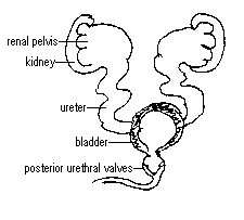 Posterior urethral valves: blockage at the outlet of the bladder
 Vesicoureteral reflux on the left: flow of urine back up ureter causing dilated ureter and kidney
 Multicystic kidney on the left: kidney may be large, leading to detection on ultrasound
 Duplication of ureters on both sides with ureterocele (seen where ureter joins bladder) on left causing bolckage
How is antenatal hydronephrosis managed?
Most cases of hydronephrosis diagnosed during pregnancy are just followed with ultrasound, monitoring the growth of the fetus and the condition of the kidneys. In these cases, a routine, normal delivery can be performed. Rarely, in a fetus with severe obstruction of both kidneys and insufficient amniotic fluid, drainage of the kidneys or bladder by tube or operation may need to be done. In these babies, however, the kidneys are often very abnormal and do not function properly regardless of treatment.
What is done to evaluate the hydronephrosis after the baby is born?
Several studies may need to be performed to evaluate the kidneys:
ultrasound (done during the newborn period)
voiding cystourethrogram (to exclude vesicoureteral reflux, a cause of 25-30% of antenatal hydronephrosis
diuretic renal scan (to evaluate kidney function)
What can be done to treat the hydronephrosis?
The treatment of antenatal hydronephrosis depends on the underlying cause. Infants and children with who have vesicoureteral reflux are managed with antibiotics and surveillance with periodic ultrasounds and voiding cystograms. Infants and children with an obstruction or blockage of the urinary tract may require surgical correction. Babies with hydronephrosis without reflux or obstruction are followed with periodic ultrasounds to monitor the hydronephrosis and the growth of the kidneys. The management of multicystic dysplastic kidneys is controversial: the multicystic dysplastic kidney doesn't work, but the opposite kidney is usually normal. Some urologists recommend removal, whereas others do not remove the dysplastic kidney unless its large size causes problems or unless there is a question of tumor or blockage.
***The information above, although based on a thorough knowledge and careful review of current medical literature, is the opinion of the doctors at Texas Pediatric Surgical Associates and is presented to inform you about surgical conditions. It is not meant to contradict any information you may receive from your personal physician and should not be used to make decisions about surgical treatment. If you have any questions about the information above or your child's care, please contact our doctors at any time by calling (713) 704-5869
You can also reach Texas Pediatric Surgical Associates by clicking HERE

Please, click HERE to go back to Kain's Main Page
Email: kainsmommy@yahoo.com

