
a.k.a. Genetics for Dummies
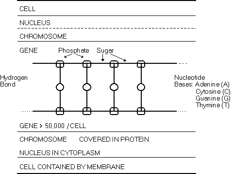
The prefix cyto- refers to the cell. The cell has a nucleus floating in cytoplasm. The cytoplasm is contained by the cell membrane. This membrane is supported by a network of filaments and tubules called the cytoskeleton which give shape and structure to the cytoplasm and allow it to move. The nucleus governs all major activities of the cell. It controls the amount and type of protein made in the cell.

The Cell | Chromosomes
| The Gene and the Protein | Muscle
Back To Info Menu
The nucleus contains 23 pairs of chromosomes, 23 maternal and 23 paternal. Chromosomes consist of long chains of nucleic acids coated with protein. There are two types of nucleic acids known as deoxyribonucleic acid (DNA) and ribonucleic acid (RNA).

DNA is the primary genetic material of all cells. DNA molecules are a long, chain-like strings of polysugar and phosphate chemical groups. DNA, which holds the coded genetic instructions and regulates enzyme production (covered later), usually consist of two intertwined chains. The chain resembles a ladder twisted into a double helix, spiral shape. Attached to each sugar in the string is a substance called a nucleotide base. The base can be one of four types. One of two purines, either adenine (A) or guanine (G) or one of two pyrimidines, thymine (T) or cytosine (C). The two chains are held together by hydrogen bonds between purines and pyrimidines (A-T and G-C).
RNA is like a single strand similar to DNA except thymine (T) is replaced by another base, uracil and the sugar-phosphate string is slightly different chemically. RNA helps transport those instructions to the cytoplasm for decoding and is generally single stranded.
The Cell | Chromosomes
| The Gene and the Protein | Muscle
Back To Info Menu
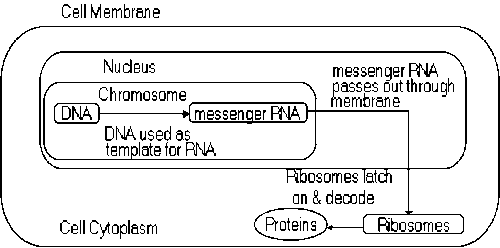
Every human cell contains more than 50,000 genes in its nucleus. Genes are segments of DNA in the chromosomes. Each gene directs the manufacture of a specific protein. Proteins have two main functions. Large, structural proteins, such as muscle fibers, are the building material of the body. Smaller proteins called enzymes regulate all cell activities. To decode a gene, a negative copy of that gene is made which is called messenger RNA. Exons are coding regions of messenger RNA which survive the processing of RNA in cell nuclei to become part of a spliced messenger of structural RNA in the cytoplasm. Currently, the protein associated with muscular dystrophy, known as dystrophin, is thought to have 79 exons.
The messenger RNA passes into the cytoplasm where it is latched on to by decoding particles called ribosomes. The decoder reads the RNA's nucleotide bases in sets of three. Each triplet of bases codes for an amino acid. Amino acids consist of nitrogenous amino and carboxyl groups of atoms linked to a variable chain or ring of carbon atoms. It is on the ribosomes where the linking of the amino acids occurs using special chemical keys known as transfer RNA. Chemical bonds, known as peptide bonds, form short chains of molecules called polypeptide chains between the carboxyl and amino groups.
Certain base triplets are also formed to terminate protein synthesis. Proteins consist of one or more polypeptide chains. The rate of protein synthesis is regulated by complex "blocking" and "unblocking" mechanisms that ensure the cell makes the right type and quantity of protein at the right time.
When healthy, the Duchenne gene carries instructions for assembling a large muscle protein known as dystrophin. Dystrophin is a large protein found on the inner side of the membrane surrounding each muscle fiber. Its purpose seems to be to maintain shape and structure of the muscle fiber similar to the way joists, rafters and posts keep a house standing. Mutations in this gene result in dystrophin deficiency which is the pathogenic basis of DMD. Dystrophin is usually absent or severely deficient in a person with DMD while those with Becker muscular dystrophy typically have dystrophin of an altered size. Without dystrophin the muscle fibers can not regenerate and are eventually replaced by fat and fiber tissue.
Now you might want to brace yourself for just one overtly technical sounding paragraph. Dystrophin has 3685 encoded amino acids which can be separated into four domains (1) N-terminal domain -240 amino acids (2) 25 triple helical segments similar to the repeat domains of spectrin (3) the cysteine-rich segment similar in part to the entire COOH domain of alpha-actin (4) C-terminal -420 amino acids. Dystrophin is predicted to be a rod shaped structural protein about 150 nanometers in length.
The Cell | Chromosomes
| The Gene and the Protein | Muscle
Back To Info Menu
Muscle is a tissue composed of of bundles of cells that have the ability to contract and relax to create movement. These cells create mechanical energy from chemical reactions. Muscles serve many functions. They produce movements of the body. They are used to maintain position of the body against gravity. They also can be used to alter pressures or tensions of structures within the body as well as protect the body. The three types or classifications of muscle are striated (skeletal), smooth and cardiac. Striated muscles attach to the skeleton. Smooth muscle is the type such as found in the stomach and blood vessels. Cardiac muscle forms the walls of the heart.
Striated muscle is the muscle of main concern for boys with DMD (although other muscles might also be affected to a lesser extent. The majority of muscles in the body are Skeletal = Striated = Voluntary muscles. They compose 40+ percent of the body weight. The term skeletal refers to the muscle's attachment to the skeleton and the ability to move bones. These muscles are under voluntary or conscious control by the brain. Striated muscles are classified by the action they perform.
|
Classification |
Action |
|---|---|
|
extensor |
opens joint out |
|
flexor |
closes joint |
|
adductor |
draws body part in |
|
abductor |
moves body part out |
|
levator |
raises body part |
|
depressor |
lowers body part |
|
constrictor |
closes orifices |
Skeletal muscle is composed of parallel groups of multi nucleate muscle fibers enclosed in a plasma membrane known as the sarcolemma. Each fiber can be thought of in segments referred to as myofibrils. The myofibrils appear as cross-banding on the fiber when viewed under a light microscope. This appearance of regular stripes leads to the term striated muscle.
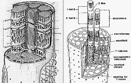
Myofibrils are enveloped in an arrangement of tubules and vesticles known as the sarcoplasmic reticulum. The sarcoplasmic reticulum is responsible for releasing and later gathering and sequestering certain chemicals such sodium, potassium and calcium ions to the myofibrils. The basic working units of the myofibril are two proteins called myosin and actin.
Each muscle fiber is supplied with a nerve ending to tell the muscle cells to contract. The brain sends an impulse to the nerve which is attached to the muscle fiber at the motor end plate. The impulse stimulates the muscle to release a neurotransmitter known as acetylcholine. This starts a chain reaction of chemical and electrical events which cause thick myosin filaments to slide over the thin actin filaments. The effect is similar to an extendable ladder being closed. Each muscle receives feedback from a set of specialized nerve fibers that register force of contraction and another set in the tendons which gage the stretch.
The Cell | Chromosomes
| The Gene and the Protein | Muscle
Back To Info Menu
