What does the word fungus bring to mind? To the farmer or homeowner,
fungi cause more than ¾ of
the plant diseases (Fig.
1-1).
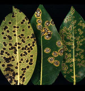
Fig. 1-1. Leaf spots caused by a fungus.
To us, we may associate fungi with ringworm, athlete's
foot
(Fig.
1-2), growth on
our culture dishes in the laboratory, or to the blackish “crud” growing in our shower stall or around the kitchen sink. After water leaks or flooding,
they can cause considerable damage to untreated wood.

Fig.
1-2.
Athlete's foot (ringworm) caused by a fungus.
Whereas, outside we see fungi as the mysterious mushrooms
(Fig. 1-3), puffballs,
and other fleshy fungi (Fig.
1-4) that appear and soon fade away. These are all fungi, but
what links them together?

Fig.
1-3. A mushroom,
Amanita muscaria.

Fig.
1-4. Bracket fungi on a tree stump.
A scientific definition for fungi is as follows:
Fungi are eukaryotic, heterotrophic
organisms that reproduce by uniflagellate
or sessile spores and whose haploid
filamentous tubular body called mycelium (Fig.
1-5)
is surrounded by walls of chitin,
glucan, rarely other compounds, and who derive their nutrition by
absorption.

Fig.
1-5. Fungal mycelium.
Included in what we call fungi are molds found in water, soil, on
foodstuff, textiles, and building materials; causing diseases of all kinds
of plants and animals; jelly fungi, bracket fungi, mushrooms, puffballs,
stinkhorns, and birdsnest fungi.
These are some confusing terms to be bouncing off
your cranium this early in the course. Let us examine each term
individually, and in that way get a better understanding about fungi. Eukaryotic
(eu=true; karyote=nucleus) organisms have true nuclei (Fig.
1-6)
and
other organelles that are membrane-bound.

Fig.
1-6. Transmission electron micrograph of a
nucleus (large membrane bound organelle) in a fungal cell.
Like other
eukaryotic organisms, fungi have nuclei with chromosomes and nucleoli.
Fungal nuclei are haploid, i.e. having a single set of chromosomes
(N), or reduced number of chromosomes in the same nucleus. Mitochondria
are membrane bound bodies that play a role in fungal metabolism (Fig.
1-7).

Fig.
1-7. Mitochondria in a fungal cell.
CLICK
HERE TO GO TO TOP OF RIGHT SIDE COLUMN
|
Fungi lack chlorophyll (achlorophyllous)
and, therefore, cannot manufacture their own food through the process of
photosynthesis. They are heterotrophic (hetero=another; trophic=food)
and must derive their nutrients from organic matter formed by other
organisms. Spores are
reproductive units usually enclosed by walls that function in
dissemination and survival. Motile spores have hair-like
structures,
flagella that
propel them, while most fungal spores are sessile, i.e. they lack flagella Spores can be of
all sizes, shapes, and colors (Fig. 1-8).

Fig.
1-8. Fungal spores of various shapes and sizes. Spores are to the fungus as seeds are to plants.
In most cases spores germinate by a germ tube and form microscopic threads
we call mycelium. A single thread is called a hypha (pl.
hyphae) (Fig. 1-9) but in most fungi the hyphae will branch extensively
to form a spawn or mold-like growth. 
Fig.
1-9. Germinated spore with hypha. The
mycelium is really the body of a fungus. When a mushroom is stimulated to reproduce, the
mycelium will differentiate and intertwine to form the button or primordial
stage of a mushroom. In real life they look like small
mushrooms (Fig. 1-10) or bracket fungi. 
Fig.
1-10. Primordial (or 'button') stage. A common mushroom will serve
to illustrate how and where spores are formed in some of our larger fungi (Fig.
1-11). 
Fig.
1-11. Mushrooms with gills.
The under surface of a mushroom bears blade-like structures we call gills.
A cross section of a gill will reveal spores formed in abundance on the
gill surface (Fig. 1-12). 
Fig.
1-12. Fungal gill with spores on the surface. A closer look with the scanning electron
microscope reveals that spores are borne on club-shaped structures called basidia (Fig.
1-13).

Fig.
1-13. Four spores borne on a basidium.
At maturity, mushrooms release spores by the millions (Fig. 1-14) and
many will land on substrates that have adequate moisture, nutrient, and
temperature to support germination.

Fig.
1-14. Mushrooms releasing millions of mature
spores.
The following image from Stamets and Chilton’s book
(The Mushroom Cultivator, Agarikon Press, Olympia, WA, 1983) depicts the
typical life cycle of a mushroom (Fig. 1-15).

Fig.
1-15. Life
cycle of a typical mushroom.
Many fungi such as the yeasts remain cellular and
their colonies consist of hundreds of individual cells. Fungal cells are
surrounded by walls containing chitin,
glucan, or rarely other compounds and are able to absorb nutrient in
liquid form. Fungal cells do not have a mouth or gullet and therefore
cannot ingest food for internal digestion. There is external digestion
and the absorption of their
solubilized nutrients.
|

