|
B. Destructive Fungal Parasites 2. Fungal Diseases of Man
|
|
|
2.
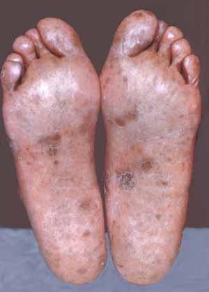
Fungal Diseases of Man: a. Superficial Mycoses: These types of mycoses are commonly referred to as dermatomycoses because they cause diseases of the skin and hair. They are caused by a number of fungi that use keratin as their basic nutrient. Tinea nigra, in North America, is caused by Cladosporium werneckii. It is a superficial, brown to black fungus infection of the epidermis, usually on palms of the hands. Black piedra forms a firmly adherent, black nodulous growth in the scalp. It occurs on various primates, including man, in humid, tropical countries. Several species of the genus Piedraia may be involved. b. Cutaneous Mycoses: This constitutes a group of fungal infections that “get under your skin”. They are caused by various species of Trichophyton, Microsporum, and Epidermophyton and in many medical mycology books goes under the name Tinea. Included among these are athelete’s foot (Fig. 13-1) which is caused predominantly by Trichophyton rubrum and ringworm of the foot caused by other species of Trichophyton and Epidermophyton floccosum.
Fig. 13-1. Athletes foot caused by Trichophyton rubrum. Species of Microsporum and Trichophyton, found commonly on cats, dogs, and other small animals, are the chief cause of ringworm of the skin. Perhaps the most common cause of ringworm of the skin (Fig. 13-2; Fig. 13-3) is Microsporum canis and Microsporum gypseum.
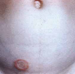
Fig. 13-2. Ringworm of the body caused by Microsporum canis.
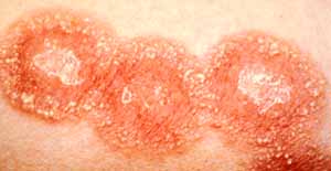
Fig. 13-3. A close up of a severe case of ringworm. Trichophyton mentagrophytes appears to be the chief cause barber’s itch, while jockey itch (Fig. 13-4; Fig. 13-5), and ringworm of the groin may be caused by yet other species of Epidermophyton and Trichophyton.
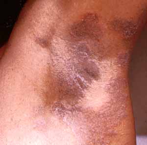
Fig. 13-4. Trichophyton rubrum causing infection of the armpit, similar to jockey itch.
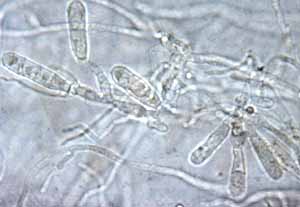
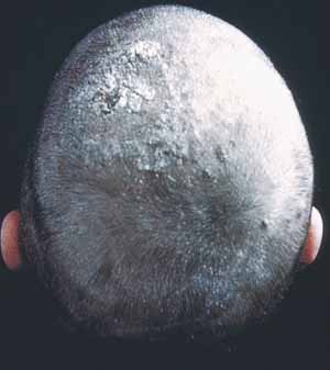
Fig. 13-5. A species Trichophyton as seen under the light microscope. Ringworm of the scalp (Fig. 13-6) is caused by various species of Microsporum and Trichophyton in which T. tonsurans seems most common.
Fig. 13-6. Ringworm of the scalp may be caused by various species of Trichophyton. Many of the dermatophytes can be grown in culture and will sporulate on keratin containing media. By using hair as bait, Microsporum and others can be isolated from soil and induced to form both its sexual and asexual (Fig. 13-7) stages. The asexual stage, Microsporium, is characterized by long, septate conidia.
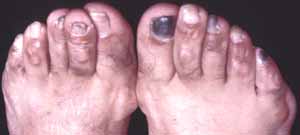
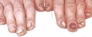
Fig. 13-7. The conidia of Microsporum canis. One of the most difficult types of cutaneous mycoses to control is the “nail fungus” caused by Trichophyton rubrum, but also by various species of Epidermophyton and Trichophyton (Fig. 13-8; Fig. 13-9).
Fig. 13-8. The world famous nail fungus, Trichophyton rubrum.
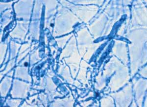
Fig. 13-9. A similar problem on fingernails. With keratin-based medium, they too can be isolated from the soil (Fig. 13-10).
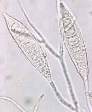
Fig. 13-10. Characteristic conidia of Trichophyton rubrum. Species of Trichophyton
have long, cylindric, multiseptate spores,
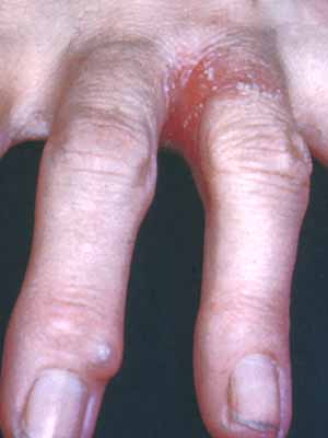
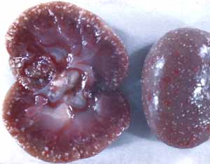
and the sexual stage consists of cleistothecia filled with globose asci. c. Subcutaneous Mycoses: These are types of mycoses that become seated in various tissues, often forming large highly inflamed, superficial tumors called mycetomas. A common cause of mycetomas is the yeast, Candida albicans that can be a problem for many who garden (Fig. 13-11). Some strains become deep-seated (see Systemic Mycoses) in the lungs and kidneys (Fig. 13-12).
Fig. 13-11. The common yeast, Candida albicans, can also get under your skin.
Fig. 13-12. The white discolored areas of the kidneys are caused by Candida albicans.
|
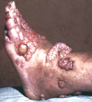
Chromomycosis is a localized chronic mycosis of skin and subcutaneous tissues. The principal causes of chromomycosis are species of Phialophora verrucosa and Cladosporium carrionii. Madura foot or maduromycosis (Fig. 13-13) is a localized swollen lesion usually on the feet or hands. It is normally caused by actinomycetes belonging to the genera Actinomyces and Streptomyces but may also be caused by certain fungal genera. In the United States, Pseudoallescheria boydii is the most common cause of Madura foot.
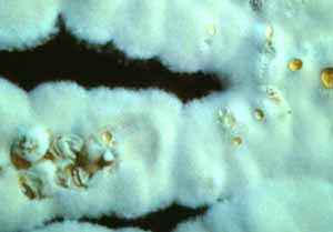
Fig. 13-13. Madura foot, caused by Pseudoallescheria boydii. A chronic, subcutaneous, lymphatic mycosis, Sporotrichosis (Fig. 13-14), may remain localized for months and then become generalized, involving bones joints, lungs and the central nervous system. This disease is caused by Sporothrix schenckii, a fungus that is widespread in various soils. This fungus forms large, globular cells within infected tissues, but becomes filamentous in culture (Fig. 13-14). Sporothrix schenckii is very common in peat moss and bags of potting mix that come from various areas of the U.S. These bags will often come with warning labels (Fig. 13-15).
Fig. 13-14. A culture of Sporothrix schenckii.
Fig. 13-15. Peat moss is a prime source of Sporothrix (bags come with warning labels). d. Systemic Mycoses: These
are types of mycoses that may involve all of the internal organs of the
body, including bones. Many of the fungi that cause systemic infections
are dimorphic, i. e. a yeast phase under some conditions and a
filamentous phase under others. Candidiasis,
mentioned earlier, is
an acute chronic, superficial or disseminated mycosis caused by species
of Candida, mostly C.
albicans. Some of the symptoms have been referred to as thrush,
endocarditis, bronchomycosis, pulmonary candidiasis, vaginitis, and
simply, yeast infection. Cryptococcosis is an acute, and sometime
chronic pulmonary, systemic mycosis caused by Cryptococcus neoformis, a
widespread budding yeast.
Pulmonary cryptococcosis is often mild and unnoticed. Cutaneous
infections, however, may become systemic and involve the central nervous
system. Geotrichosis
is an oral, intestinal, bronchial, or pulmonary infection caused by Geotrichum
candidum. It is usually a secondary infection in which the fungus is
an opportunistic pathogen. It often causes chronic or acute bronchitis,
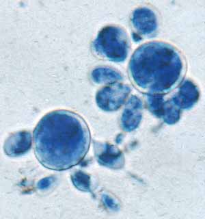
tracheitis, or pulmonary lesions. Perhaps one of the most important systemic mycoses is Blastomycosis, caused by Ajellomyces (Blastomyces) dermatidis, a dimorphic fungus that grows in mammalian tissues as budding cells (Fig. 13-16), but on culture media as a dry white fungal growth.
Fig. 13-16. Ajellomyces dermatidis is the cause of blastomycosis. Blastomycosis is a chronic granulomatous disease that originates as a respiratory infection. A. dermatidis has often been isolated from the soil and is felt to survive on organic debris within the soil. Lobo’s disease (Fig. 13-17), also referred to as South American blastomycosis, caused by Paracoccidioides brasiliensis, is a chronic, often fatal mycosis characterized by primary pulmonary lesions that may disseminate to many visceral organs. It is endemic to several areas in the U.S.
Fig. 13-17 Lobo's disease is caused by Paracoccidioides brasiliensis. Coccidioidomycosis, also called valley fever or desert rheumatism, is a benign, severe or sometimes fatal mycosis. It is respiratory in origin and may be limited to the upper respiratory tract and lungs. It may spread to visceral organs, bones, and joints, or form burrowing abscesses in the skin (Fig. 13-18).
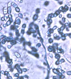
Fig. 13-18. Coccidioidomycosis is a type of mycetoma caused by Coccidioides immitis. It is caused by Coccidioides immitis, a species that forms large yeast cells within infected tissues but becomes filamentous in culture (Fig. 13-19). The disease is endemic in the desert areas of southern Califormia, Arizona, New Mexico, southwestern Texas, and various Central and South American countries, and has been shown to be more prevalent after rain.
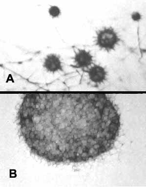
Fig. 13-19. Coccidioides immitis as seen in culture. Histoplasmosis is an intracellular mycosis of the reticuloendothelial system involving lymphatic tissue, lungs, spleen, liver, kidneys, skin and central nervous system. It is caused by the dimorphic fungus Histoplasma capsulatum that is able to survive in soil that is enriched with the excrement of bats or birds. It is found in caves, around poultry houses, and under roosting areas of native birds. It has been shown to be a keratinophilous fungus whose ascal state belongs in Gymnoascus (Fig. 13-20), one of the Plectomycetes we studied earlier. The asexual state, H. capsulatum, is easily recognized in culture by its globose spores with peg-like ornaments (Fig. 13-20).
Fig. 13-20. A cleistothecium of Gymnoascus (B), and it asexual stage, Histoplasma capsulatum (A). e. Miscellaneous Mycoses: Several
genera of fungi with numerous species may be responsible for mycotic
infections in man. Among them includes: Aspergillosis,
a granulomatous disease of lungs, but occasionally spreads to other
organs. Aspergillus fumigatus is the usual causal agent of pulmonary
aspergillosis. Other species of Aspergillus
and other unrelated fungi occasionally are found in this type of
infection. Less common is a mycosis referred to as Penicillosis
caused by a number of species of Penicillium
and Scopulariopsis. While
these fungi may be opportunistic
pathogens on inflammed mucus membranes of the eye, ear, nose, and
throat, they often cause pulmonary mycosis. A number of fungi with dark
pigments cause what is referred to as phaeohyphomycosis
(phaeo=pigmented; hyphomycetes=condial fungi). One of the most common of
these is Cladosporium.
Phaeohyphomycosis and many other types of mycoses are carried by animals. |