|
B. Destructive Fungal Parasites 3. Fungal Diseases of Domesticated Animals
|
|
|
3. Fungal Diseases of Domesticated Animals Not
surprising since we are mammals, most of the fungal diseases of man are
found on domesticated animals. They
are probably all present on wild mammals, but we are not aware of many
because they have not been studied. Domesticated animals have superficial
mycoses, subcutaneous mycoses, and systemic mycoses.
There are a number of conditions that are conducive to fungal infections
in animals in tropical and subtropical countries. They include hot, humid
climates, overcrowding, and unsanitary conditions. Transmission of such
diseases may be by direct contact, or by air-borne or water-borne spores.
There are a number of clinical signs that signify a possible fungal
infection. These include broken hairs, scales or crusts, papules, or
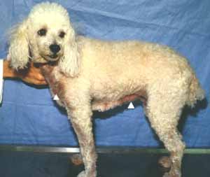
pustules. However, some animals are asymptomatic. a. Superficial mycoses include Malassezia pachydermatis that is caused by Pityrosporum canis, and is common on dogs in north central Florida (Fig. 14-1).
Fig. 14-1. Malassezia pachydermatis (arrows) is the most important superficial disease on dogs. This fungus causes what has been termed tinea versicolor or pityriasis in man, in which superficial white, brown or fawn colored, superficial lesions occur. Their sexual states belong to cleistothecial members of the Plectomycetes. Piedra, a disease as in man, gets on the skin and hair shafts of animals, producing a crustose surface. Fungi that are involved in superficial mycoses include species of Microsporium and Trichophyton, in fact, the same species that are found on man, for example, Trichophyton equinum (Fig. 14-2) and Microsporum nanum (Fig. 14-3). While species of these genera occur on a variety of animals, they appear more prevalent on dogs and cats.
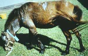
Fig. 14-2. Superficial mycoses on horses may be caused by Trichophyton equinum.
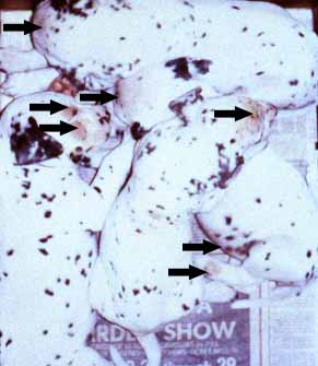
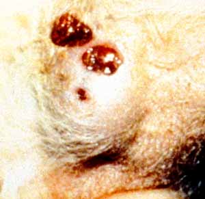
b. Subcutaneous infections such as sporotrichosis also occur in animals (Fig. 14-5; Fig. 14-6). It is caused by Sporothrix schenckii (Fig. 14-4), a widespread saprophyte in the soil .
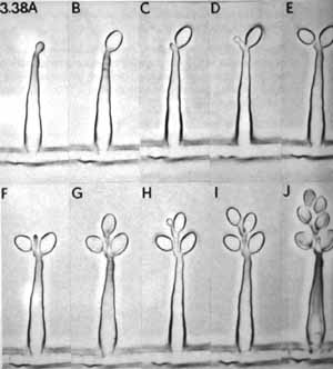
Fig. 14-4. The microscopic mature of Sporothrix schenckii.
Fig. 14-5. Puppies showing symptoms of sporotrichosis (arrows).
Fig. 14-6. Cats can also be susceptible to sporotrichosis.
|
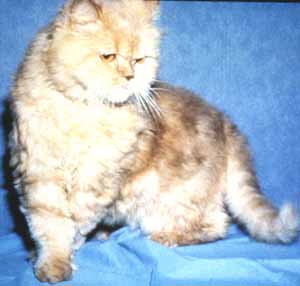
Many animals, especially long-haired cats may have superficial mycoses and never show symptoms. They are called asymptomatic carriers (Fig. 14-7).
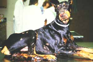
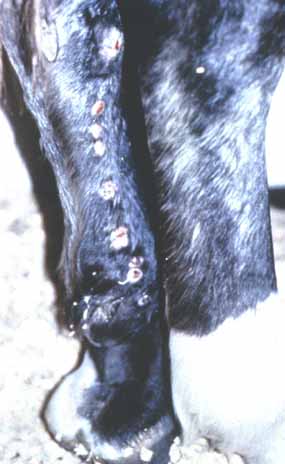
c. Systemic infections include mycetomas, phaeohyphomycosis, chromoblastomycosis, rhinospopridiosis, and phytiosis. Some rather severe systemic infections may occur in animals that spend considerable time standing in water, especially certain variety of dogs (Fig. 14-8) and horses (Fig. 14-9).
Fig. 14-8. Symptoms of phytiosis caused by Pythium insidiosum.
Fig. 14-9. Phytiosis is also a common problem on horses. Of
importance among these is Pythiosis caused by species of the water mold Pythium, the same group that causes damping off in plants!
Many of these may start out as subcutaneous lesions, and if left
untreated, cause gastrointestinal diseases.
During the drought of 2000 and early 2001, many of the lakes in Florida
were extremely low. Lake sediment that was high in nutrient was being
dredged and used on lawns, gardens, and pastures as a good organic
fertilizer. Unfortunately, previously submerged plant material was
infested with Pythium insidiosum that causes large
gastrointestinal masses which if left untreated becomes lethal.
Domesticated animals also contract aspergillosis,
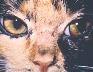
penicilliosis, and candidiasis. Widespread systemic mycoses include blastomycosis, histoplasmosis, and coccidioidomycosis. Many of these are endemic in their geographical distribution. Histoplasmosis is associated with bats and birds, and their symptoms are much like those in man, pulmonary, lymphatic, and hematogenous. Coccidioidomycosis, or San Joaquin Valley Fever, is common in the Southwestern US deserts in drought conditions followed by rain. It may start as an eye infection but quickly develops into deep-seated mycosis (Fig. 14-10; Fig. 14-11).
Fig. 14-10. Symptoms of coccidioidomycosis on the eye of a dog.
Fig. 14-11. Mycetomas are also frequently associated with coccidioidomycosis. The yeast, Cryptococcus neoformis, also causes Cryptococcosis in animals in which white lymphomas may occur. Several different fungi, many opportunistic pathogens, may cause mycetomas. Mycetomas caused by pigmented genera of Hyphomycetes are called phaeohyphomycosis. They include genera such as Curvularia, Dreschlera, Cladosporium, and Phialophora. (Images of mycoses of domesticated animals were provided by the staff of the small animal clinic, Univ. of Florida Veterinary College)
|