Endocrinology Topics ![]()
![]()
 Endocrine
Pancreas
Endocrine
Pancreas
The pancreas ("Pan" = all; "creas" = flesh) is composed of both exocrine acinar cells and endocrine cells grouped in Islets of Langerhans. Exocrine functions of the pancreas described in the 17th Century, endocrine functions in the 19th Century.
Paul Langerhans, a German medical student, described unusual clusters of cells in pancreas - the “Islets of Langerhans” in 1869. In 1893, Laguesse suggested the islet cells produce a substance that may prevent diabetes mellitus. In the same year, Joseph von Mering & Oscar Minkowski showed that dogs got diabetes after removal of the pancreas.
By 1905, Eugene Gley in France showed that pancreatic extracts cured diabetes in dogs. But this escentric scientist did not publish his results but they were sealed in a packet and deposited with the French Society of Biology to be opened later. Nicolas Paulesco in Rumania also showed that pancreatic extracts cured diabetes in dogs in 1916 and publishe a paper in August 1921.
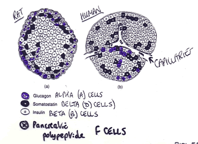 Islets
have at least four types of endocrine cells: alpha (A),
beta (B), delta (D) and
F cells. Alpha cells produce glucagon. Delta cells produce somatostatin. Beta
cells produce insulin and amylin. F cells produce pancreatic polypeptide.
Islets
have at least four types of endocrine cells: alpha (A),
beta (B), delta (D) and
F cells. Alpha cells produce glucagon. Delta cells produce somatostatin. Beta
cells produce insulin and amylin. F cells produce pancreatic polypeptide.
Since it is a mixed endocrine and exocrine gland, the pancreas develops from dorsal and ventral buds that evaginate from the duodenum at aboyt embryonic day 9 in mice, mostly from endodermal origins (Gittes and Rutter, PNAS USA 89:1129-1132 ; 1992). Even before the pancreas is formed, the primitive gut produces somatostatin by embryonic day 8. By dau 9 the primitive pancreatic buds can produce insulin and glucagon. By day 10 the pancreatic buds produce pancreatic polypeptide and by day 10.5 carboxypeptidase A. Buds fusing and the acini forms by day 12, and amylase is produced.
Insulin Promoter Factor 1 (IPF1 aka STF-1 or IDX-1), a homeodomain transcription factor regulation pancreatic development. IPF1 activates the insulin gene promoter, is selectively expressed in the beta cells of adult pancreas and is first seen in the region of the primitive gut that commits to a pancreas fate. Homozygous IPF1 gene knockouts mice survive fetal development but lack the pancreas and die a few days after birth. Also have no ectopic expression of insulin or pancreatic enzymes.
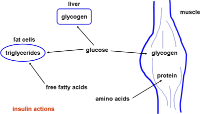
 Insulin regulates
storage of fuel and has growth properties. It facilitates uptake of glucose
by the muscle, liver and adiposites, amino acid uptake by the muscle and fat
uptake by adipocytes.
Insulin regulates
storage of fuel and has growth properties. It facilitates uptake of glucose
by the muscle, liver and adiposites, amino acid uptake by the muscle and fat
uptake by adipocytes.
Proinsulin gives rise to A and B chains of insulin (21 and 30 aa, respectively). Insulin circulates unbound as a monomer. It is almost completely reabsorbed by proximal tubules of kidney and degraded by the kidneys. The liver removes about half of the insulin that passes through it.
 Insulin
signals by binding a monomeric tyrosine kinase membrane receptor capable of
autophosphorylation. The active receptor then activates the cytosolic proteins
IRS1/2 and SHC, which can then interact with a variety of proteins through their
SH2 or SH3 domains. IRS-1 and IRS-2 are different and may regulate different
pathways or stages or condition. Activation of the SH2/3 proteins leads to activation
of the kinases MAPK and AKT, and translocation of the glucose transporter GLUT4
to the membrane. Both MAPK and AKT signaling lead to glycogen synthase activation
(liver and muscle) while GLUT4 increases glucose uptake. MAPK also leads to
gene transcription of target genes.
Insulin
signals by binding a monomeric tyrosine kinase membrane receptor capable of
autophosphorylation. The active receptor then activates the cytosolic proteins
IRS1/2 and SHC, which can then interact with a variety of proteins through their
SH2 or SH3 domains. IRS-1 and IRS-2 are different and may regulate different
pathways or stages or condition. Activation of the SH2/3 proteins leads to activation
of the kinases MAPK and AKT, and translocation of the glucose transporter GLUT4
to the membrane. Both MAPK and AKT signaling lead to glycogen synthase activation
(liver and muscle) while GLUT4 increases glucose uptake. MAPK also leads to
gene transcription of target genes.
Insulin receptor knockout mice (D. Accili et al., Nature Genetics 12:106, 1996) are born at term with normal intrauterine growth and development, but have hyperglycaemia and hyperketonaemia and die as result of diabetic ketoacidosis 48-72 hrs after birth. This is consistent with a model in which the insulin receptor primarily mediates the metabolic actions of insulin. A similar human genetic disorder caused by mutations in the insulin receptor is known as "leprachaunism": severe intrauterine growth retardation, dysmorphic features and disturbed glucose metabolism.
IRS-1 mice knockouts have retarded growth and have a mild resistance to insulin, but do not get diabetes due to increased insulin secretion. IRS-1 may also mediate some effects of IGF-1 and IGF-2.
IRS-2 in mice knockouts have impaired insulin signaling and B cell function. They suffer a progressive deterioration of glucose metabolism due to insulin resistance in liver and skeletal muscle. This suggests that IRS-2 dysfunction may be important in type 2 diabetes.
<insert about Caenorhabditis elegans and DAF-16 later>
 The
glucagon receptor increases intracellular cAMP levels which activates a signaling
cascade that culminates in activation of phsphorylase a, the enzymes that calalyzes
the first step in the glycogenolitic pathway.
The
glucagon receptor increases intracellular cAMP levels which activates a signaling
cascade that culminates in activation of phsphorylase a, the enzymes that calalyzes
the first step in the glycogenolitic pathway.
Somatostatin is made by D cells of the gut, pancreas and hypothalamus. In the pancrease, somatostatin is co-secreted with insulin. Somatostatin inhibits glucagon and insulin release, but its exact role is unclear. Pancreatic somatostatin may restrain entry of nutrients into the body by inhibiting digestive/hormonal events.
Pancreatic polypeptide is made by F cells of pancreas, stimulated by hypoglycemia or a protein meal. It decreases liver glycogen, stimulates lipogenesis, inhibits gallbladder contraction and pancreatic enzyme secretion in humans. Pancreatic polypeptide may be inhibited by somatostatin.
Amylin is a human islet 37 amino acid polypeptide (hIAPP), major component of insoluble amyloid deposits in NIDDM patients. It is made by B cells of pancreas (like insulin) but also acts as a neuroendocrine hormone (?). The amylin receptor is the calcitonin receptor with a helper protein called a RAMP.
Amylin is co-secreted with insulin after a meal, inhibits glucagon secretion, slows gastric emptying and lowers effective glucose intake. It appears that for carbohydrates the rate of absorption in the intestine is linked to the rate of delivery from the stomach (i.e. gastric emptying rate). Thus amylin slows the delivery rate and therefore the glucose absorption rate. This allows insulin to do its job better and the patient can achieve better sugar control. A synthetic version (drug) of amylin is Pramlintide (Amylin Inc.).
Adiponectin (aka complement-related protein 30 [ACRP30], apM1, adipoQ) is a 30 kDa protein produced by adipocytes (not a pancreas-produced hormone) involved in regulation of lipid and carbohydrate metabolism. It enhances insulin action in animals, lowers glucose levels without stimulating insulin secretion, lower glucose levels in patients with type 2 diabetes, obese patients and patients with insulin resistance. May be protective against cardiovascular disease.
Take Quiz: [Q1] [Q2] [Q3]
Back to Basics: Topic 1
Topic 2
Advance Topics: Topic 1
Topic 2
Insulin and Metabolism
Carbohydrate metabolism of the liver includes several pathways: glycolysis (glucose --> Pyruvate --> ATP), glycogen breakdown (phosphorylase driven?), pentose phosphate pathway (produces reducing power), gluconeogenesis (from non-carbohydrate precursors. also in kidney) and glycogen synthesis (polymeric reaction driven by glycogen synthase). Many of these pathways are regulated by insulin and glucagon. [Synthases are enzymes that catalyze condensation reactions in which no nucleotide triphosphate is required as an energy source. Synthetases are enzymes that catalyze condensation reactions using ATP or another nucleoside triphosphate as an energy source.]
 Intestinal
L cells secrete glucagon-like peptide (GLP-1) in response to nutrient ingestion.
GLP-1 induces insulin biosynthesis and secretion, as well as islet neogenesis
and prevention of B cell destruction.
Intestinal
L cells secrete glucagon-like peptide (GLP-1) in response to nutrient ingestion.
GLP-1 induces insulin biosynthesis and secretion, as well as islet neogenesis
and prevention of B cell destruction.
Pancreatic B cells secrete insulin when blood glucose levels rise. Insulin levels are increased by: raised blood glucose or amino acids, glucagon, gastrin, secretin, GLP-1, cholecystokinin (CCK), gastric inhibitory peptide (GIP), alpha-adrenergic sympathetic innervation and parasympathetic innervation. Insulin is decreased by low blood glucose, somatostatin, beta-adrenergic sympathetic innervation and stress.
Pancreatic A cells secrete glucagon in response to low blood glucose. Glucagon produces the biological actions opposite to insulin: promotes hepatic gluconeogenesis, lipolysis and glycogenolysis. It is a 29 amino acid polypeptide made by the A cells from a 179 amino acid preproglucagon molecule. Glucagon is similar in structure to VIP, secretin and GIP. Glucagon is stimulated by hypoglycemia, CCK, VIP, and arginine. It is inhibited by free fatty acids, insulin, secretin, somatostatin, and hyperglycemia.
During the catabolic phase of carbohydrate metabolism (4-6 hours after food intake) the body uses endogenous fuels and glucagon higher. In the adipose tissue, fatty acids are converted to ketones, that normally are used as fuel outside the CNS (except during starvation). The muscle converts proteins to glucose and the liver breaks down glycogen stores into glucose, which can be used by the CNS.
Glycogen synthase, responsible for the formation of glycogen, is heavily regulated by pancreatic hormones. Insulin inhibits the kinase that activates glycogen synthase by no-cAMP signaling. Glucagon stimulates the same kinase via cAMP signaling. Insulin stimulates the phosphatase that dectivates glycogen synthase.
 Glucose
is the major fuel source for the brain (~120 g per day). About 60% of body glucose
is used by the brain. During starvation, the brain uses ketones intead. Free
fatty acids cannot be used as fuel source becasue they are albumin-bound and
cannot pass the blood/brain barrier. In addition, brain tissue has no glucose-6-phosphatase.
Glucose
is the major fuel source for the brain (~120 g per day). About 60% of body glucose
is used by the brain. During starvation, the brain uses ketones intead. Free
fatty acids cannot be used as fuel source becasue they are albumin-bound and
cannot pass the blood/brain barrier. In addition, brain tissue has no glucose-6-phosphatase.
Muscle uses glucose, fatty acids and ketones as fuel and has glycogen stores (75% of body stores are in muscle). But muscle has no glucose-6-phosphatase therefore cannot export glucose. The muscle keeps glucose stores for bursts of activity (glycolysis) and exports metabolized pyruvate to the liver as lactate. Fatty acids are a major fuel source of resting muscle.
Triacylglycerols are major fuel source in adipose tissue. Glucose is needed from liver for triacylglycerides synthesis. Adipose tissue breaks triacylglycerides down to glycerol and fatty acids using lipases.
The liver provides fuel for brain, muscle and adipose (glucose by gluconeogenesis). When fuels are abundant, the liver makes fatty acids which are then esterified and sent to the blood as very low density lipoproteins (VLDLs). During fasting, fatty acids are converted to ketones.
Take Quiz: [Q1] [Q2] [Q3]
Back to Basics: Topic 1
Topic 2
Advance Topics: Topic 1
Topic 2
Diabetes Mellitus
Ancient descriptions of diabetes mellitus can be found in Chinese, Hindu and Greek writings. “Diabetes” is Greek for “to pass through” and “mellitus” means “sweet-tasting” (sugar in urine). In the 18th century, Dobson in England described high blood and urine sugar in diabetics. In December 2nd, 1921, Leonard Thompson, a 13 year old boy, was admitted to Toronto General Hospital with juvenile-onset diabetes mellitus. He would have probably died soon if Banting and Best had not injected him with an extract from beef pancreas glands. By the 1980s recombinant DNA techniques allowed synthesis of pure human insulin in bacteria. Banting and Best (working in the MacLeod lab in Toronto) described their purification of insulin in 1922. The 1923 Nobel Prize was awarded to Banting and MacLeod. The 1958 Nobel Prize was awarded to Fred Sanger of Cambridge University "for his work on the structure of proteins, especially that of insulin".
14 to 16 million people in the US have diabetes mellitus, up to 50% of cases going undiagnosed. In the US, more people die annually from diabetes than from AIDS, breast cancer, and automobile accidents combined. Diabetes is a major cause of death and dissability in the US. It occurs equally in males and females. There are three types: insulin-dependent (IDDM, aka Type 1 or juvenile onset), non-insulin dependent (NIDDM, aka Type 2 or adult onset), and gestational. Type 1 accounts for 5-10% of cases, while Type 2 accounts for 90-95%. Gestational may be temporary or develop into Type 2.
Type 1 diabetes is due to auto-immune disease that destroys the B cells of the pancrease. The sorce of the auto-immune disease is not known, but may involve genetic or viral factors. It is more common in whites than non-whites. Symptoms include increased thrist and urination, constant hunger, weigth loss and blurred vision. Glucose will build up in the blood and if untreated will lead to diabetic coma. Gestational diabetes develops or is discovered during pregnance. It often disappears after pregnancy, but patients are more likely to later get Type 2. Type 1 and gestational diabetes are successfully managed by a daily regimen of insulin injections.
 Type
2 diabetes is more common in non-whites, and native americans have specially
high rates. The onset of the disease occurs around age 40 and is more common
in overweight patients. Very obese patients may even get type 2 diabetes early
in life (chilhood-teens). The pancreas initially produces adequate insulin but
the peripheral tissues cannot use it efficiently. Thus there is a high production
of insulin in order to compensate, but this eventually fails, leading to diabetic
symptoms that develop gradually: feeling unwell, increased thirst and urination,
wegth loss, blurred vision, infections, and slow-healing of skin injuries.
Type
2 diabetes is more common in non-whites, and native americans have specially
high rates. The onset of the disease occurs around age 40 and is more common
in overweight patients. Very obese patients may even get type 2 diabetes early
in life (chilhood-teens). The pancreas initially produces adequate insulin but
the peripheral tissues cannot use it efficiently. Thus there is a high production
of insulin in order to compensate, but this eventually fails, leading to diabetic
symptoms that develop gradually: feeling unwell, increased thirst and urination,
wegth loss, blurred vision, infections, and slow-healing of skin injuries.
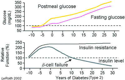
Loss of action by the normal amount of insulin (i.e. insulin resistance) may exist for a decade before onset of Type 2 diabetes. This is associated with obesity and may be mediated by an increase in free fatty acids. The compensatory hyperinsulinemia eventually leads to B cell dysfunction, declining insulin secretion and hyperglycemia.
Complications due to diabetes involve both microvascular and macrovascular damage: blindness, heart disease, stroke, kidney failure, amputations and nerve damage. Patients often first become aware they have diabetes after developing one of these life-threatening complications. Diabetes is diagnosed by one or more of the following tests:
Fasting glucose levels of 110-125 mg/dL (6.1-6.9 mM) are a sign of Impaired Glucose Tolerance (IGT), a condition which is likely to progress to diabetes. In patients with IGT, diet and excersise reduce the risk of progressing to diabetes.
Glucose-stimulated insulin secretion is defective in the diabetic pancreas, and with time appropriate insuline secretion fails. A possible new drug target to stimulate insulin secretion is recombinant GLP-1 or a GLP-1 analog like exendin-4 (Exenatide), a GLP-1 agonist isolated from the saliva of the Gila monster lizard with a longer t1/2 than GLP-1. In clinical trials, exendin-4 controlled patient blood sugar and reduced their weight. Another alternative is to decrease GLP-1 clearance (t1/2 = 1-2 min) by inhibiting dipeptidase IV.
There is unrestricted glucose production in the diabetic liver, through increased rates of gluconeogenesis and glycogenolysis (processes normally inhibited by insulin). The defective insulin-driven processes in adipose tissue are still unclear.
In the muscle, glucose is not taken up efficiently due to insulin resistance. A new drug target to enhance insulin action in the muscle are a small-molecule activators or inhibitors, for example protein tyrosine phosphatase 1b (PTP-1b) inhibitors. PTP-1b is an ubiquitously expressed tyrosine phosphatase that blocks insulin-stimulated activation of the insulin receptor. Mice deficient in PTP-1b exhibit increased insulin sensitivity as well as resistance to weight gain when fed a high-fat diet.
Since Type 2 diabetes is more of an organ resistance to insulin, no hormone injections are required in most cases, but other pharmacological agents are used to increase the insulin sesitivity of target organs. Oral medications for treating Type 2 diabetes include sulfonylureas, biguanides, alpha glucosidase inhibitors, and PPARg ligands. They can be used alone or in combination according to the needs of the patient.
Sulfonylureas stimulate insulin production in the pancreas. Unfortunatelly they may accelerate the exhaustion of B cells and become inefective over time. First generation sulfonylureas include chlorpropamine (Diabinase), and Orinase. Second generation are used in lower doses, thus are safe, and include glyburide (Micronase, Diabeta) glipizide (Glucotrol XL), repaglinide (Prandin), nateglinide (Starlix), and glibenclamide (Euglucon). Because the stimulate insulin production, hypoglycemia is a concern. They are taken with meals.
Biguanides like glucophage (Metformin) reduce glucose release from the liver. Since they do not stimulate insulin secretion hypoglycemia is not a side effect. Alpha glucoside inhibitors like acarbose (Precose) and miglitol (Glyset) delay degestion and absorption of sugar into de blood. Alpha-glucosidase is an intestinal enzyme required for carbohydrate digestion. Taken before meals, the alpha-glucoside inhibitors partially block carbohydrate absorption, thus lowering peak glucose levels after meals and controlling postprandial blood glucose levels.
The PPARg ligands are also known as thiazolidinediones (TZD), glitazones or insulin sensitizers, and include drugs like troglitazone (Rezulin), rosigitazone (Avandia) and piogitazone (Actos). When used in combination with insulin or sulfonilureas may cause hypoglycemia. Troglitazone was pulled from the market after causing several deaths due to liver failure. TZDs target insulin resistance by increasing peripheral tissue sensitivity to insulin. PPARg is predominantly expressed in adipose tissue and TZDs mediate changes in adipose gene expression. TZDs increase expression of adiponectin, and decrease expression of TNFa and resistin (adipocyte-derived cytokine, causes insulin resistance and glucose intolerance). This has insulin-sensitizing effects in muscle and liver, but patients often gain weight.
Current diabetes research focuses in several areas: genetics, improving blood glucose management, improving insulin delivery technology, and treatments for peripheral tissue damage. Identifying the genes involved in Type 1 and/or Type 2 diabetes should help in the development of new drugs and therapies. Intensive management of blood glucose helps prevent or delay the onset of complications, including the use of better non-invasive blood glucose monitors, excersise, weigth management and diet. Development of anti-obesity agents that reduce appetite and/or increase energy expendituyre will also lead to effective treratment and prevention of Type 2 diabetes.
Non-invasive nasal sprays, pills, or pathches, as well as implantable insulin pumps can replace daily injections. Transplantation of donor or artificial B cells is not yet available. Laser treatment could be used for diabetic eye disease. Antihypertensive drugs may prevent or delay kidney failure.
Take Quiz: [Q1] [Q2] [Q3]
Back to Basics: Topic 1
Topic 2
Advance Topics: Topic 1
Topic 2
![]()
![]() Continue
to "Gonadal Development" or take
a test: [T1] [T2] [T3].
Continue
to "Gonadal Development" or take
a test: [T1] [T2] [T3].
Need more practice? Answer the following review questions:
1- What are Islets of Langerhans?
Clusters of endocine cells in the pancreas.
2- List 4 types of pancreatic endocrine cells and their products,
Alpha - glucagon
Beta - insulin and amylin
Delta - somatostatin
F cells - pancreatic polipeptide
3- Describe the developmental origin of the pancreas.
Develops from dorsal and ventral buds that invaginate from the duodenum, mostly
from endodermal origins.
4- In what order are pancreatic hormones available during development?
Somatostatin - before pancreas formation because there are D cells in the primitive
gut
Insulin and glucagon - from primitive pancreatic buds
P ancreatic pollipeptide - from later buds
5- What is Insulin Promoter Factor 1 (IPF-1)?
Homeodomain transcription factor needed for pancreatic development. Activates
the insulin gene promoter, is selectivelly expressed in B cells of the adult
pancreas, and is first seen in the region of the primitive gut that commits
to a pancreasfate.
6- What is the phenotype of homozygous IPF-1 mice knockouts?
Survive fetal development but lack the pancreas and die a few days after birth.
7- What are the general functions of insulin?
Regulates storage of fuel and has growth-inducing properties. Facilitates uptake
of glucose by the muscle, liver and adipocytes, amino acid uptake by muscle
and fat uptake by adipocytes.
8- Descrbe the general structure of insulin.
Proinsulin gives rise to A and B chains joined by bisulfide links.
9- What is the fate of insulin in the circulation?
Circulates unbound as a monomer, is almost completely reabsorbed by the proximal
tubules of the kidneys and also degraded in the kidneys. The liver removes about
half that passes through.
10- Briefly describe the insulin receptor.
Monomeric tyrosine kinas membrane receptor capable of autophosphorylation.
11- What are the IRSs?
Cytosolic phosphotyrosine proteins activated by the insulin receptor, which
when active interact with a variety of cytosolic proteins through their SH2/3
domains. IRS-1 and IRS-2 are different and may regulate different pathways or
stages or conditions.
12- List 4 protein kinases activated by insuling signaling.
PI3 kinase
AKT
MAPK
S6 kinase
13- List 3 transcription factors activated by insulin signaling.
JUN
FOS
MYC
14- Which enzyme involved in carbohydrate metabolism is activated by insulin
signaling?
glycogen synthase
15- Briefly describe the glucagon signaling pathway.
The glucagon receptor increases intracellular cAMP, which activate a signaling
cascade that culminates in activation of phosphorylase a, the enzyme that catalyzes
the firs step in the glycogenolitic pathway.
16- Were is somatostatin made?
D cells of the gut, pancreas and hypothalamus. In the pancreas is cosecreted
with insulin.
17- What are the functions of somatostatin regardin carbohydrate metabolism?
Inhibits insulin ang glucagon release, but its exact role is unclear, may restrain
entry of nutrients into the body. May inhibit pancreatic polypeptide.
18- Were is pancreatic polypeptide made and by what stimuli?
in F cells of the pancreas, stimulated by hypoglycemia or a protein meal
19- What are the functions of pancreatic polypeptide?
Decreases liver glycogen, stimulates lipogenesis, inhibits gallblader contractions
and pancreatic enzyme secretion.
20- What is amylin?
Major component of insoluble amyloid deposits in NIDDM patients, made by B cells
and co-secreted with insulin.
21- What is the function of amylin?
Inhibits glucagon secretion and slows gastrict emptying, thus allowing insulin
to do a better job, lowering the effective glucose intake .
22- What is the amylin receptor?
Acts as a a neuroendocrine hormone, its receptor is the calcitonin receptor
with a helper protein, RAMP.
23- What is adiponectin?
Hormone produced by adipocytes involved in regulation of lipid and carbohydrate
metabolism.
24- What are the effects of adiponectin?
Enhances insulin action, lowers glucose levels witout stimulating insulin secretion,
lowers glucose levels in patients with type 1 diabetes, insulin rewsistance
or obesity, may be protective against cardiovascular disease.
Remining questions not yet available