Endocrinology Topics
![]()
![]()
 Anatomy
and Development
Anatomy
and Development
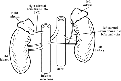
The adrenals are two glands located over each of the kidneys that mediate the body's short and long term responses to stress. They have a very good blood suply. The right adrenal drains to the inferior vena cava, whil the left adrenal drains to the left renal vein. The glands have two structuraly different areas: the medulla and the cortex.
The adrenal medulla is made of cromaffin cells that produce cathecholamines, which are stored in granules until signalled to release. Cromaffin cells are oxidized by potassium dichromate into a brown color, thus their name. Each chromaffin cell is inervated by a synpathetic neuron, therefore the adrenal medulla could be considered a modified ganglion.

The cells in the cortex make steroids, are rich in lipid droplets, and are arranged into three distint structures: zona glomerulosa, zona fascicilata and zona reticularis. The zona glomerulosa is arrended into ovoid groups of cubidal cells. The zona faciculata is arranged into cords of cells with spongy appearance. The zona reticularis is an interconnecting network of cells and sinusoidal blood vessels.
The adrenal medula and cortex have different embrionic origins. The adrenal medula develps from the neural crest ectoderm, from the same orign as the sympathetic and parasympathetic ganglia. The fetal medula makes noradrenaline, but only epinephrine after birth. The adrenal cortex develps from visceral mesoderm.
While mamals have adrenal glands composed of medulla and cortex, other animals may have the ectodermal an mesodermal tissues iseparated or in different conformations. In reptiles the medula is on one side of the cortex. In birds and anphibians the medullary tissue is dispersed inside the cortex tissue, and in amphibians the whole adrenal is enveloped by kidney tissue. In sharks the medullary tissue is compleatelly outside the cortex tissue and dispersed through a larger anatomical area.
Take Quiz: [Q1] [Q2] [Q3]
Back to Basics: Topic 1
Topic 2
Advance Topics: Topic 1
Topic 2
 Adrenal
Medula
Adrenal
Medula
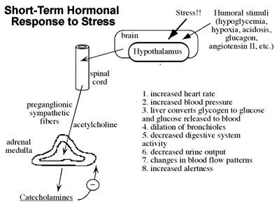
The adrenal medula mediates short term responses to stress and humoral stimuli transmited via neural signaling to the medula.
The chromaffin cells of the medulla produces the cathecholamines epinephrine (adrenaline) and norepinephrine (noradrenaline). Cathecholamines increase heart rate and blood pressure, induce conversion of glycogen to glucose in the liver and the release of glucose to the bloodstream, dilate bronchioles, change blood flow patterns, increase alertness and decrease the activity of the digestive system and urine output.
A given chromaffin cell usually secretes either adrenaline or noradrenaline. Other proteins in granules include the chromogranins, ex. chromogranin A (acidic). About 80% of the hormone produced by the medulla is epinephrine. Noradrenaline is also a neurotransmitter eleased by postsynaptic (postganglionic?) neurons of the sympathetic nervous system (SNS).
Catecholamines are made from tyrosine produced by the liver. The rate limiting step is the conversion of tyrosine to dopa by tyrosine hydroxylase in the cromaffin cell's cytoplasm. Dopa is converted to dopamine, which in turn is collected by granules.
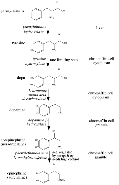 Two
more enzymatic steps occur inside the granules in order to convert dopamine
into epinephrine. The enzyme phenylethanolamine-N-methyltransferase
catalyzes the last step, the conversion of norepinephrine to epinephrine. This
methyltransferase is negativelly regulated by epinephrine and norepinephrine
(negative feedback) and induced by high glucocorticoid levels.
Two
more enzymatic steps occur inside the granules in order to convert dopamine
into epinephrine. The enzyme phenylethanolamine-N-methyltransferase
catalyzes the last step, the conversion of norepinephrine to epinephrine. This
methyltransferase is negativelly regulated by epinephrine and norepinephrine
(negative feedback) and induced by high glucocorticoid levels.
As hormones, epinephrine and norepinephrine have some similar physiological effects, but in many cases they have opposite effects. Both epinephrine and norepinephrine increase blood pressure respiration, metabolism, and dilation of blood vessels in muscle. But heart rate and skin blood vessel dilation are increased by epinephrine and decreased by norepinephrine. Cardiac output increases with epinephrine but the effects of norepinephrine are variable. Epinephrine facilitates passive/tense behaviour while norepinephrine facilitates agressive behaviour. Epinephrine also increases eosinophil count.
| Effect | Epinephrine | Norepinephrine |
| Heart rate | increases | decreases |
| Cardiac output | increases | variable |
| Blood pressure | increases | increases |
| Respiration | increases | increases |
| Metabolism | increases | small increase |
| Behavior | passive, tense | aggressive |
| Eosinophil count | increases | ---- |
| Skin blood vessels | dilate | constrict |
| Muscle blood vessels | dilate | small dilation |
Epinephrine and norepinephrine
signal through several types of adrenergic receptors, all are 7-transmembrane,
G-protein coupled receptors. b1and b2
adrenergic receptors signal by stimulating G-proteins (Gas)
to activate the cAMP pathway. a1 adrenergic receptors signal through a Gq
protein to activate the IP3/CA![]()
![]() pathway. a2
adrenergic receptors signal through an inhibiory G-protein (Gai)
to block the cAMP pathway.
pathway. a2
adrenergic receptors signal through an inhibiory G-protein (Gai)
to block the cAMP pathway.
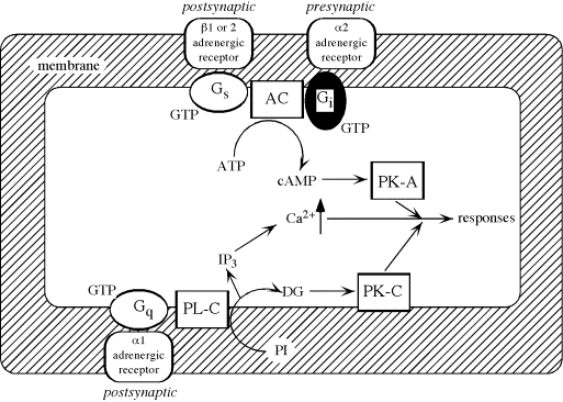
a2 receptors located in the presynaptic neuron signal to decrease the release of norepinephrine into the synaptic celft (negative feedback). Even in the presence of constant norepinephrine in the synaptic cleft, response decreaseas due to receptor desensitization: receptors are internalized and inactivated by phosphorylation.
Epinephrine and norepinephrine activate a1 and a2 receptors more or less equally. a-adrenergic receptors receptors mediate vasoconstriction and uterine muscle contraction, amoung other physiological effects. Phenylephrine is a syntetic a-adrenergic receptor agonist. Phentolamine is a syntetic a-adrenergic receptor antagonist.
Epinephrine is a better ligand for the b1and b2 receptors than norepinephrine. b-adrenergic receptors mediate cardiac stimulationm and vasodilation. Isoproterenol is a syntetic b-adrenergic receptor agonist, and is a stronger ligand than epinephrine. Other b-adrenergic receptor agonists include caffeine and the amphetamines. Propanolol is a syntetic b-adrenergic receptor antagonist.
Catecholamines have very short half-lives (10-20 seconds). They are metabolized by monoamine oxidase in most tissue, and by cathecol-o-methyltransferase in the liver and kidney. 95% of epinephrine and norepinephrine is excreted in urine as their metabolite vanillic acid, only 5% is excreted intact.
Take Quiz: [Q1] [Q2] [Q3]
Back to Basics: Topic 1
Topic 2
Advance Topics: Topic 1
Topic 2
 Adrenal
Cortex
Adrenal
Cortex
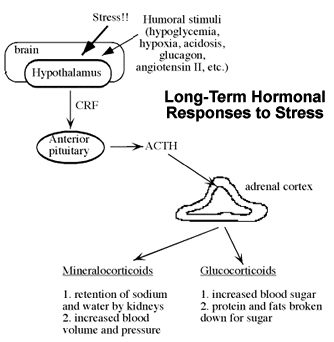
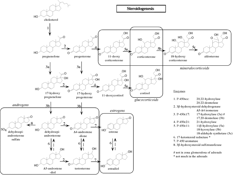
The adrenal cortex responds to stress and humoral stimuli transmited via ACTH stimulation from the anterior pituitary. The cortex produces three types of steroids: mineralocorticoids, glucocorticoids and androgens. Steroids have an 18 carbon, 4 ring structure and are synthesized from cholesterol. The zona fasticulata (cortisol) and zona reticularis (DHEA) are the major ACTH-responsive layers of the adrenal cortex. Hypophysectomy leads to atrophy of this leyers.
The mineralocorticoids affect water balance and are made in the zona glumerulosa. The glucocorticoids affect metabolism and the immune system, and are made in the zona fasciculata. The androgens do what? and are made in the zona reticularis.
Lampreys and similar fishes have only traces of steroid hormones. Sharks and bony fishes have glucocorticoids only. Lungfishes have aldosterone and cortisol. All tetrapods have aldosterone. Amphibians, reptiles and birds also have glucocorticoids, although different chemicals predominate in the different species.
Which steroid is made in which cells depends on the enzymes present. The first step of steroid synthesis is the conversion of cholesterol into pregnenolone by P450sec. The key enzyme for entrance into the androgenic metabolic pathway is P450c17, which is not present in great quantity in the areas were mineralocorticoids or glucocorticoids are made. The enzyme that converts androstenedione into testosterone is not abundant in the adrenal cortex.
Take Quiz: [Q1] [Q2] [Q3]
Back to Basics: Topic 1
Topic 2
Advance Topics: Topic 1
Topic 2
Glucocorticoids
Cortisol is made in the zona fasticulata (intermediate layer). It is the major glucocorticoid in humans and other primates, while corticosterone is the main glucocorticoid in rodents. Cortisol release is episodic and variable, following ACTH pulses by about 15-30 mins. Then it excerts negative feedback on CRH and ACTH release. There is a burst of cortisol release just before waking up in the morning. About 80% of the cortisol in the bloodstream is bound to transcortin (GC binding alpha 2-globulin). About 15% is bound to albumin. The half-life of cortisol is 70 - 90 minutes.
Cortisol is essential for life: it maintains critical processes during prolonged stress and contains inflammation. Most effects of cortisol are permissive, many organs have cortisol receptors:
| Liver | Gluconeogenesis to increase glycogen stores |
| Fat | Lipolysis to free fatty acids |
| Muscle | Proteolysis - atrophy |
| Parturition | Fetal cortisol initiates parturition in sheep |
| Connective tissue | Inhibits growth |
| Immune/ Suppression | atrophy, anti-inflammatory |
| Water metabolism | Promotes water retention |
| Pituitary | Inhibits ACTH release from anterior pituitary |
| CNS | Modulates perception and emotion |
| Development | Intestine, adrenal medulla, lungs (surfactant?) |
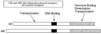
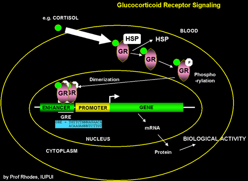 The
glucocorticoid receptor (GR) is a nuclear receptor, i.e. a ligand-dependent
transcription factor part of the GR/MR family and part of the nuclear receptor
superfamily). Nuclear receptors exist in the cytoplasma bound to chaperones
(ex. HSP) or maybe bound to DNA in the nucleus but with little transactivation
activity, until bound by their ligand. Receptor phosphorylation may also increase
activity. The active receptor then dimerizes, binds DNA, and regulates transcription
of genes with a hormone response element (HRE) in ther pormoter, i.e. a GRE
in the case of GR.
The
glucocorticoid receptor (GR) is a nuclear receptor, i.e. a ligand-dependent
transcription factor part of the GR/MR family and part of the nuclear receptor
superfamily). Nuclear receptors exist in the cytoplasma bound to chaperones
(ex. HSP) or maybe bound to DNA in the nucleus but with little transactivation
activity, until bound by their ligand. Receptor phosphorylation may also increase
activity. The active receptor then dimerizes, binds DNA, and regulates transcription
of genes with a hormone response element (HRE) in ther pormoter, i.e. a GRE
in the case of GR.
Targeted disruption of the glucocorticoid receptor gene blocks adrenergic chromaffin cell development and severely retards lung maturation (Cole et al.; Genes & Dev. 9: 1608-1621; 1995). GR knockout mice (homozygous) die a few hours after birth of respiratory failure. Lung development is impaired (atelectatic?). Liver gluconeogenesis enzyme genes are down-regulated. Feedback is lost therefore ACTH and corticosterone are high, and the adrenals are enlarged (cortical hypertrophy) .There is little or no medulla and no epinephrine.
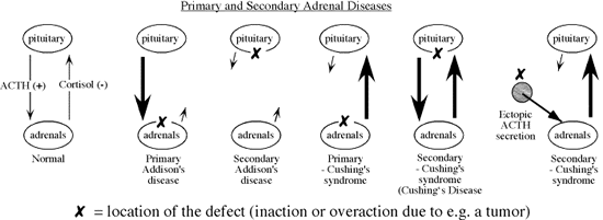
The main diseases of cortisol malfunction are Cushing's syndrome and Addison's disease. Cushing's Syndrome is due to overproduction of cortisol. Symptoms include obesity, fatigue, high blood pressure, bruising, female menstrual problems, moon face, and buffalo hump. Cushing's be caused by pituitary adenomas that secrete ACTH, by other tumors that secrete ACTH, or by adrenal tumors that secrete cortisol. WHn caused by pituitary adenomas that secrete ACTH it is knwon as Cushing's Disease.
Addison's Disease (AD) is a rare condition of adrenal failure. Primary AD is usually due to tuberculosis, fungal infections, or autoimmune disease. Secondary AD is caused by insufficient ACTH. The major symptoms are low blood pressure and fainting. In primary AD, ACTH levels can get very high (no negative feedback at the anterior pituitary) and hyperpigmentation may occur.
Take Quiz: [Q1] [Q2] [Q3]
Back to Basics: Topic 1
Topic 2
Advance Topics: Topic 1
Topic 2
 Mineralocorticoids
Aldosterone is made in the zona glomerulosa (outermost layer). The mineralocorticoid receptor (MR) binds aldosterone and glucocorticoids with equal affinity. In aldosterone target tissues, the MR is protected from glucocorticoids by 11b-hydroxysteroid dehydrogenase type 2 (11b-OHD2). Thus inactivation of glucocorticoids allows aldosterone to specifically activate the MR in eipithelial cells of the distal colon and the prpincipal cells of the collecting ducts in hte kidney. Howver, in MR-expressing cells that lack 11b-OHD2, MR is maily activated by glucocorticoids, ex: neurons of the limbic system.
MR knocout mice die in the second week after birth of hypoaldosteronism, and activity of the ENaC (?) is impaired in kidney. The MR knockout mice can be kept alive with a salt solution. (see Berger et al., Kidney Int. 57: 1295, 2000). MRKO mice have a normal prenatal development but during the first week of postnatal life develop pseudohypoaldosteronism. At day 8 they show hyperkalemia, hyponatremia, and a strong increase in renin, angiotensin II and aldosterone plasma concentrations. They loose weight and die around day 10 from dehydration by renal loss of sodium and water. Thesodium excretion was elevated 8-fold. MRKO neonates die because they are not able to to compensate renal sodium loss. Regulation of sodium reabsorption via MR is not achieved by transcriptional control of ENaC and Na/K-ATPase in RNA abundance but by transcriptional control of other as yet unidentified genes. (?) Daily subcutaneous injections of isotonic NaCl solution until weaning and continued oral NaCl supply lead to survival of MRKO mice. However, the renal salt-loss defect persist.
Renin/Angiotensin/Aldosterone - a kidney/adrenal axis. Drop in blood volume/pressure => macula densa sodium levels change => transmural pressure changes => renal nerve signal. Juxtaglomerular cells of glomerulus produce renin hormone. Renin acts on angiotensinogen leading to the production of angiotensins I, II and III. Angiotensin II promotes aldosterone production and causes sodium influx to the kidney => blood volume rises. Angiotensin II also stimulates thirst, ADH release from pituitary, cardiac output and peripheral resistance => blood volume and pressure rises.
<more>
DHEA is made in the zona reticularis (innermost layer). <more later>
Take Quiz: [Q1] [Q2] [Q3]
Back to Basics: Topic 1
Topic 2
Advance Topics: Topic 1
Topic 2
Subtitle
Text
Take Quiz: [Q1] [Q2] [Q3]
Back to Basics: Topic 1
Topic 2
Advance Topics: Topic 1
Topic 2
Subtitle
Text
Take Quiz: [Q1] [Q2] [Q3]
Back to Basics: Topic 1
Topic 2
Advance Topics: Topic 1
Topic 2
![]()
![]() Continue
to "nest topic" or take a test: [T1] [T2] [T3].
Continue
to "nest topic" or take a test: [T1] [T2] [T3].
Need more practice? Answer the following review questions:
Questions not yet available