Digestive System
Introduction
Single-celled organisms can directly take in nutrients from their outside environment. Multicellular animals, with most of their cells removed from contact directly with the outside environment, have developed specialized structures for obtaining and breaking
down their food. Animals depend on two processes: feeding and digestion. Animals are heterotrophs, they must absorb nutrients or ingest food sources. The digestive system uses mechanical and chemical methods to break food down into nutrient molecules that can be absorbed into the blood. There are two types of plans and two locations of digestion. Sac-like plans are found in many invertebrates, who have a single opening for food intake and the discharge of wastes. Vertebrates use the more efficient tube-within-a-tube plan with food entering through one opening (the mouth) and wastes leaving through another (the anus).
Intracellular digestion: food is taken into cells by phagocytosis with digestive enzymes being secreted into the phagocytic vesicles; occurs in sponges, coelenterates and most protozoans.
Extracellular digestion: digestion occurs in the lumen (opening) of the digestive system, with the nutrient molecules being transferred to the blood or body fluid; occurs in chordates, annelids, and crustaceans.
Stages in the Digestive Process:
1.movement: propels food through the digestive system
2.secretion: release of digestive juices in response to a specific stimulus
3.digestion: breakdown of food into molecular components small enough to cross the plasma membrane.
4.absorption: passage of the molecules into the body's interior and their passage throughout the body
5.elimination: removal of undigested food and wastes
Too often we are inclined to think that proteins and carbohydrates nourish us but it is actually the amino acids and sugars that actually enter our blood and nourish our cells.
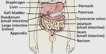
The human digestive system is a coiled, muscular tube (6-9 meters long when fully extended) extending from the mouth to the anus. Several specialized compartments occur along this length: mouth, pharynx, esophagus, stomach, small intestine, large intestine, and anus. Accessory digestive organs are connected to the main system by a series of ducts: salivary glands, parts of the pancreas, and the liver and gall bladder. The digestion of food requires a cooperative effort between different parts of the body.
Identify and give a function for each of the following:
- mouth
- Ingestive eaters, the majority of animals, use a mouth to ingest food. In humans the digestion of starch also begins here. [Absorptive feeders, such as tapeworms, live in a digestive system of another animal and absorb nutrients from that animal directly through their body wall]- tongue and teeth -
Mechanical breakdown begins in the mouth by chewing (teeth) and actions of the tongue. [The tongue is composed of striated muscle with an outer layer of mucous membrane]. The tongue manipulates food during chewing and swallowing. It mixes food with saliva and then forms the mixture into a bolus in preparation for swallowing. Mammals have tastebuds clustered on their tongues. Taste buds play a role in what we call taste but it is largely a product of the olfactory receptors in the nose. 20 deciduous teeth. 32 adult teeth. 4 types: incisors (biting), canine (tearing), premolars (grinding), molars (crushing)- salivary glands -
Three pairs of exocrine glands (the parotid, sublingual, and submandibular glands) that send their juices by way of ducts to the mouth. Chemical breakdown of starch by production of salivary amylase from the salivary glands. Mucus moistens food and lubricates the esophagus. Bicarbonate ions in saliva neutralize the acids in foods. This mixture of food and saliva is then pushed into the pharynx and esophagus. [Also contains lysozyme (antibacterial) and antibodies][ Mumps begins as infective parotitis in the parotid glands in the cheek]- pharynx
- Swallowing moves food from the mouth through the pharynx into the esophagus and then to the stomach.- epiglottis
- The opening from the pharynx to the trachea (the glottis) is covered during swallowing by a flap of tissue called the epiglottis. Fig 11.4 page 175- esophagus -
The esophagus is a muscular tube leading from the pharynx to the stomach whose muscular contractions (peristalsis) propel food to the stomach.- cardiac sphincter
- constrictor muscle at the entrance of the stomach that acts as a valve. Relaxes during swallowing. Failure ----> heartburn. When vomiting occurs a reverse peristaltic wave causes the sphincter to relax and the contents of the stomach are propelled upward through the esophagus.
- cardiac sphincter
- constrictor muscle at the entrance of the stomach that acts as a valve. Relaxes during swallowing. Failure ----> heartburn. When vomiting occurs a reverse peristaltic wave causes the sphincter to relax and the contents of the stomach are propelled upward through the esophagus.- stomach
- During a meal, the stomach gradually fills to a capacity of 1 liter, from an empty capacity of 50-100 milliliters. At a price of discomfort, the stomach can distend to hold 2 liters or more. Epithelial cells line inner surface of the stomach, and secrete about 2 liters of gastric juices per day. Gastric juice contains hydrochloric acid, pepsinogen, and mucus; ingredients important in digestion. Secretions are controlled by nervous (smells,thoughts, and caffeine) and endocrine signals. Hydrochloric acid (HCl) lowers pH of the stomach so pepsin is activated (and pH kills most bacteria). Pepsin is an enzyme that controls the hydrolysis of proteins into peptides. Mucus protests the wall of the stomach from HCl. Failure ---> Ulcer [However, there is now evidence that a bacterial infection by Heliobacter pylori may impair the ability of cells to produce protective mucous] The stomach also mechanically churns the food. Chyme, the mix of acid and food in the stomach, leaves the stomach and enters the small intestine.
- pyloric sphincter -
The pyloric sphincter repeatedly opens and closes, allowing the chyme to enter the small intestine in small squirts only. [stomach empties in 2 - 6 hours.]- duodenum
The first 25 cm of the small intestine where ducts from the gallbladder and pancreas join and enter the duodenum- liver and gall bladder -
Bile, a watery greenish fluid is produced by the liver and secreted via the hepatic duct and cystic duct to the gall bladder for storage, and thence on demand via the common bile duct to an opening near the pancreatic duct in the duodenum. It contains bile salts, bile pigments (mainly bilerubin, essentially the non-iron part of hemoglobin) cholesterol and phospholipids. Bile salts and phospholipids emulsify fats, the rest are just being excreted. [Gallstones are usually cholesterol based, may block the hepatic or common bile ducts causing pain, jaundice.]- pancreas -
produces pancreatic juice which contains sodium bicarbonate (basic) and neutralizes the acidity of the chyme as it enters the duodenum. [Endocrine and exocrine gland. Exocrine part produces many enzymes which enter the duodenum via the pancreatic duct. Endocrine part produces insulin, blood sugar regulator]- small intestine -
Receives bile from the gall bladder and secretions from the pancreas, chemically breaks down chyme, absorbs nutrient molecules, and transports undigested material to the large intestine.- large intestine (colon) -
The large intestine is made up by the colon, cecum, appendix, and rectum. Material in the large intestine is mostly indigestible residue and liquid. Movements are due to involuntary contractions that shuffle contents back and forth and propulsive contractions that move material through the large intestine. Secretions in the large intestine are an alkaline mucus that protects epithelial tissues and neutralizes acids produced by bacterial metabolism. Water, salts, and vitamins are absorbed, the remaining contents in the lumen form feces (mostly cellulose, bacteria, bilirubin). [Bacteria in the large intestine, such as E. coli, produce vitamins (including vitamin K) that are absorbed.]
- appendix -
The appendix is a small projection on the large intestine near the entrance of the small intestine. In humans the appendix may play a role in immunity. [This organ is subject to inflammation a condition called appendicitis. It removal is delayed and the appendix burst it can lead to a generalized infection of the abdominal cavity.]- rectum -
The last 20 cm of the large intestine.- anus -
The rectum opens at the anus where defecation, (expulsion of feces) occurs. Feces contains nondigestible remains, bile pigments (give color), and bacteria (give smell).Relate the following digestive enzymes to their glandular sources and describe the digestive reactions they promote:
- salivary amylase -
component of saliva produced by the salivary glands. Begins the process of digesting food, specifically starch. With the addition of water the enzyme allows starch to be converted to maltose.- pancreatic amylase - produced by the pancreas and sent to the duodenum. With the addition of water the enzyme allows starch to be converted to maltose
- proteases (pepsin, trypsin) - With the addition of water these enzymes allow protein to be converted to peptides. Pepsin is produced and used in the stomach. Trypsin is produced by the pancreas and sent to the duodenum.
- lipase - produced by the pancreas and sent to the duodenum. With the addition of water the enzyme allows fat droplets to be converted to glycerol + 3 fatty acids. [Before lipase can act the fat has to be emulsified into fat droplets by the bile salts.]
- peptidase - present in the mucosa of the intestinal villi of the small intestine, peptidase completes the digestion of peptides into amino acids (with the addition of water).
[only after this reaction are the molecules small enough to cross the cell membrane]
- maltase- present in the mucosa of the intestinal villi of the small intestine, maltase completes the digestion of maltose into glucose (with the addition of water). [only after this reaction are the molecules small enough to cross the cell membrane]
[Notice that the digestion of starch and proteins is a two step process]
- nuclease - A team of enzymes called nucleases produced in the small intestine and pancreas, hydrolyze DNA and RNA in food into their component nucleotides. Other hydrolytic enzymes then break nucleotides down further into nitrogenous bases, sugars, and phosphates.
Note: There will be a test question on figure 11.13 and very likely an exam question based on a similar experiment.
Describe swallowing and peristalsis
Swallowing moves food from the mouth through the pharynx into the esophagus and then to the stomach.
 Step 1: A mass of chewed, moistened food, a bolus, is moved to the back of the moth by the tongue (voluntary). In the pharynx, the bolus triggers an involuntary swallowing reflex that prevents food from entering the lungs, and directs the bolus into the esophagus.
Step 2: Muscles in the esophagus propel the bolus by waves of involuntary muscular contractions (peristalsis) of smooth muscle lining the esophagus. (Fig. 11.9 Page 179)
Step 3: The bolus passes through the gastroesophageal sphincter, into the stomach. Heartburn results from irritation of the esophagus by gastric juices that leak through this sphincter.
Identify the components and describe the digestive actions of gastric juice
Gastric juice contains HCl and pepsin. The HCl does not digest food but it does break down the connective tissue in meat and provides the low pH that pepsin needs to operate. Pepsin digests proteins into peptides.
Identify the components and describe the digestive actions of pancreatic juice
 (a)Amylase Acts on starch
Starch--> Maltose
(b)Trypsin Acts on proteins
Proteins --> Amino acids
(c)Lipase Acts on fats (lipids)
Fats --> Glycerol + 3 fatty acids
(d)Sodium bicarbonate Buffer, neutralizes acid chyme
Identify the components and describe the digestive actions of intestinal juice
Peptidase - Acts on peptides thus completing the digestion of protein into amino acids. Peptides + H2O ---> amino acids
Maltase - Act on maltose thus completing the digestion of starch into glucose
Maltose + H2O ---> glucose
Identify the source gland for and describe the function of insulin
The endocrine portion of the pancreas called the islets of Langerhans produces and secretes insulin [and glucagon] directly into the blood. All the cells of the body use glucose as an energy source. It is important that the glucose concentration remain within normal limits. Insulin is secreted when there is a high level of glucose in the blood, which usually occurs just after eating. Insulin has three different actions.
(1) it stimulates the liver, fat and muscle cells to take up and metabolize glucose
(2) it stimulates the liver and muscle to store glucose as glycogen
(3) it promotes the buildup of fats and proteins and inhibits their use as an energy source
[Glucagon stimulates the breakdown of stored nutrients and causes the blood glucose level to rise.]
Explain the role of bile in the emulsification of fats

List six major functions of the liver

1) detoxification of blood;
2) synthesis of blood proteins;
3) destruction of old erythrocytes and conversion of hemoglobin into a component of bile;
4) production of bile;
5) storage of glucose as glycogen; and
6) production of urea from amino groups and ammonia.
[Glycogen (chains of glucose molecules) serves as a reservoir for glucose. Low glucose levels in the blood cause release of hormones to stimulate breakdown of glycogen into glucose. When no glucose or glycogen is available, amino acids are converted into glucose in the liver. The process of deamination removes the amino groups from amino acids. Urea is formed and passed through the blood to the kidney for export from the body.]
[Jaundice occurs when the characteristic yellow tint to the skin is caused by excess hemoglobin breakdown products in the blood, a sign that the liver is not properly functioning. Jaundice may occur when liver function has been impaired by obstruction of the bile duct and by damage caused by hepatitis. Hepatitis A, B, and C can all cause liver damage. Cirrhosis of the liver commonly occurs in alcoholics, who place the liver in a stress situation due to the amount of alcohol to be broken down.]
Examine the small intestine and describe how it is specialized for digestion and absorption
- The wall of the small intestine contains fingerlike projections called villi that increase the total absorptive surface area. Each villus is further subdivided into a series of yet smaller villi (microvilli) which increase the absorptive surface area even more. [The lining of the small intestine as a result of these wrinkles upon wrinkles has a total absorptive surface area of 600 square meters, about the size of a baseball diamond.] Each villus contains blood vessels and a small lymphatic vessel called a lacteal. [fig. 11.7 page 177] The lymphatic system is an adjunct to the circulatory system; its vessels carry a fluid called lymph to the circulatory veins. The microvilli bear the intestinal digestive enzymes which finish the digestion of chyme to molecules small enough to cross the membrane of a cell. [The intestinal digestive enzymes are also called brush-border enzymes because the microvilli that give the interior of the small intestine a fuzzy border in electron micrographs.]
Describe the functions of E. coli in the colon
Bacteria in the large intestine, such as E. coli, produce vitamins (including vitamin B and K) that are absorbed. Bacteria also produce characteristic gasses which may stimulate defecation reflexes.