TEAM
4.2 |
||
|
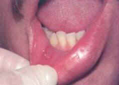
Aphthous ulcers, also known as canker sores or aphthous stomatitis (2), occur on areas of the mouth in which the mucosa is nonkeratinised and loosely attached, particularly the buccal mucosa, labial mucosa, floor of the mouth, ventral surface of the tongue, and soft palate (1).
Recurrent aphthous ulcers usually occur in recurrent bouts at intervals of a few days to a few months (6).They may appear initially as erythematous indurated papules that erode to form sharply circumscribed necrotic ulcers covered by a thin grey fibrinous exudate and rimmed by a narrow zone of erythema. These ulcers can be differentiated from viral or bacterial infections and similar dermatologic conditions by the lack of distinguishing systemic features and the healthy appearance of adjacent tissue (1). There are three clinical variations of RAU. All are believed to be part of the same disease spectrum, and correspond to the degree of severity (i.e. size and number of ulcers) (4) 1. Minor aphthous ulcers: Occur in 80-85% of RAU cases (1) and are usually less than 5 mm in diameter. Most commonly, one to five ulcers are present at any one time and they usually heal spontaneously (without scarring) within 7-10 days (3).
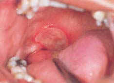
2. Major aphthous ulcers (also termed Sutton disease): Approximately 10-15% of RAU cases (1). These ulcers are larger (>10 mm in diameter) and deeper, tend to have irregular edges, and are more painful. They can take 10-30+ days to heal, and are often accompanied by considerable scarring (3).
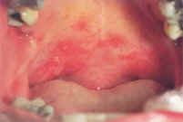
3.Herpetiform RAUs: Occur in 5-10% of RAU cases (1). These are 1 to 3 mm in diameter and often occur in groups of more than 100, which often coalesce to form large, irregular areas of ulceration. These are not as deep as major RAUs and heal without scarring in 7 to 14 days. In spite of their name, they are not of viral origin or associated with herpes virus (3).
In most cases, diagnosis of aphthous ulcers can be deduced from history and the characteristic of clinical response (12). There are no laboratory procedures or medical testing to confirm the diagnosis (11.13). If there is any doubt about the diagnosis then measures should be taken to exclude other possible causes of oral ulceration. Patients with persistent and troublesome RAS should undergo screening such as full blood count and film, and measurement of inflammatory markers and haematinic (serum ferritin, serum B12, serum and red cell folate) to identify any underlying haematinic deficiency (14).
It is estimated that at least 1 in 5 individuals has at least once been afflicted with aphthous ulcers (2). Prevalence may be increased in affluent countries and socio-economic classes. A study of medical and dental students revealed a prevalence of 31-66% (1). Aphthous ulcers may be slightly more common in females than in males. Outbreaks occur more frequently during ovulation or before menstruation, and remissions are common during pregnancy (1) The peak age of RAU onset is 10-19 years, with a tendency to decrease in severity and frequency with age (5). Major aphthous ulcers may begin soon after puberty whereas herpetiform RAUs tends to affect older persons (1).
Risk factors for RAUs include (10):
Three stages are recognized in the development of an aphthous ulcer (8).
The pathophysiology of RAUs remains incompletely understood and they are often termed idiopathic. They appear to be multifactorial in origin, with a strong component being immune mediated (3). Several contributing factors have been proposed(4):
Some of these possibilities will now be discussed in more detail. Patient with severe aphthous ulcer is noted in 60% of those with immunodeficiencies. These patient lacks one more immunoglobulin classes (low serum IgG, IgM, IgA) due to B-cell abnormalities. IgA deficiency is a common primary immunodeficiency and it was speculated that the IgA deficiency mechanism is connected to an immunoregulatory defect in a form of suppressor T-cell overactivity. 4. Microbiologicviral, bacterial Streptococcus sanguis has been suggested as important in the pathogenesis of RAU (and Behcets disease), either as direct pathogens or an antigenic stimulus, culminating in the genesis of antibodies that cross-react with keratinocyte antigenic determinants, but this theory was discarded (4,5). The clinical similarity of oral aphthous ulcers to secondary herpes simplex virus (HSV) infections lead to an investigation of a viral cause of RAU. However, this also was not substantiated (4).
Genetic factors are likely to predispose patients to RAUs, and more than 40% of affected individuals have first-degree relatives with RAS(9). RAU may be associated with the human leukocyte antigen (HLA) haplotypes B51 (also common in Behçet syndrome), Cn7, A2, B12, and Dr5 (1).
A number of precipitating factors to RAU have been implicated. These include nutritional deficiencies, and malabsorption (1). Other causes of aphthous ulcers that have been investigated include emotional stress, food hypersensitivity, hormonal imbalance, systemic disorders and trauma. Although none of these factors have been found to be important in the primary causation of aphthous ulcers, they may have a modifying or triggering role(4) Deficiencies of iron, folic acid, zinc, and vitamins B1, B2, B6, B12 have all been implicated in RAU (1). Although deficiencies were identified in only a small percentage of patients, correction of these deficiencies produced improvement or cures in this all of the group (4). Malabsorption conditions such as celiac disease, ulcerative colitis and Crohn's disease have been associated with aphthous-type ulcers (1,4). However, this should be treated with a high degree of suspicion since the ulcers may be the only presenting symptom for a number of years in patients with GI tract disorders. Also, deficiencies of folic acid and factors related to the underlying disease may be part of the cause of RAUs in these patients. The role of psychological and physiologic stress as risk factors for aphthous ulcers is controversial (1). Individuals with aphthous ulcers have been noted to have higher-than-average anxiety scores and cortisol levels (1). Although there is no convincing evidence linking hypersensitivity to foods with RAUs, some patients recognize that foods ( such as chocolate, cheese and tomatoes) and certain flavouring agents and essential oils precipitate attacks of RAS (5). In some women, RAU is associated with the menstrual cycle, outbreaks are more common during ovulation or before menstruation (1). Some patients report being free from aphthae whilst taking oral contraceptives or when pregnant (10). Smoking and nicotine exposure do not increase, and may actually decrease, the risk of aphthous ulcers1. Some patients report the onset of oral ulcers after smoking cessation. Also, nicotine-containing tablets appear to control the frequency of RAS (7). Patients with systemic disorders, such as cyclic neutropenia, Reiter syndrome, Behçet disease or HIV may have more severe and protracted aphthous-like ulcers, however the exact role of these disorders is unknown (1,4).
To prevent occurrence or recurrence of aphthous ulcer, patients are advised to avoid trigger factors such as food with sharp edge, brushing teeth too hard (which injure teeth, gum and oral mucosa), or reducing stress in life (adopting relaxation technique). It is also recommended that patients keep a mouth ulcer diary. It is a simple note book which keep track of the toothpaste patient use, the food they eat, stressful events, and others including recording any trauma that may occur to the inside of their mouth. Patients are also required to keep record of the number of the aphthous ulcer occurrence. This would allow patients to identify the specific trigger factor causing the ulcer.
| |||||||||||||||||||||
| ||||||||||||||||||||||