Viruses
Viruses and bacteria are too small to be seen without the aid of microscopes. As disease agents, their effects on mankind are well known. Both are ubiquitous and adaptable.
The Bridge Between Living and Non-Living
Someone once suggested that if people were the size of viruses, the entire population of the U.S. would fit on the end of two pencil erasers. There would be room left over for future generations. Extremely small, simple in structure, and widely distributed, viruses exist in a realm all their own. Viruses do not qualify as cells yet affect cells and so exist as if on a bridge between the living and nonliving.
Structure and Classification of Viruses
Viruses differ from cellular organisms in many ways. A virus
contains only a single type of nucleic acid. This DNA or RNA may be single or double-stranded. The core of nucleic acid is covered by a protein coat called a capsid. Some of the proteins in the capsid are enzymes. A complete virus particle is called a virion. Some viruses have an additional outer covering or envelope. This envelope comes from the cell membrane of the cell infected by the virus. The capsid and envelope protect the virus from physical damage. They prevent chemical damage by enzymes in the host cell. Some of these enzymes, called nucleases, break down nucleic acids.
The outer covering of the virus may contain spike-like extensions or filaments. Some viruses even have a tail made of protein. These extensions help the virus to bind to the surface of specific cells. This binding is necessary for the virus to infect the cell. The type of nucleic acid, the structure of the outer covering and dispersal mechanisms are features used to classify viruses. Viruses infect all forms of life. Organisms and physical objects that carry or transmit viruses are called vectors. Viruses that infect bacteria are called bacteriophages, or simply phages. Phages are some of the most studied viruses.
More than 1000 plant diseases are caused by viruses. Tomatoes, potatoes, and cucumbers are some of the plants affected. Damage to the cell wall of a plant cell allows plant viruses to enter. Plant viruses may be transferred by contaminated machinery, fungi, nematode worms, and sucking insects like aphids. Even pollen and seeds may transmit viruses.
Animal and human viruses are among the best known. Measles, influenza, smallpox, Herpes, and AIDS are just a few examples. Few of us would forget to put the common cold into this group. Animal viruses attach to the plasma membrane of an animal cell. They enter the cell by phagocytosis or pinocytosis. Enveloped viruses may enter by fusing their envelope with the plasma membrane. The entire plant or animal virus enters the host cell. A bacteriophage injects its nucleic acid into the bacterial cell.The capsid remains outside.
Most viruses can only be seen with an electron microscope.
Viruses lack the structures we associate with cells. Thus, they are called subcellular, noncellular, or acellular infectious agents. Viruses must use the metabolic machinery of a live host cell to produce more viral particles. A virus has no metabolism of its own. It reproduces only after taking over a living cell. Thus, a virus is considered an INTRACELLULAR, OBLIGATE, PARASITE. It is a protected particle of nucleic acid. It depends upon a living cell to carry out the functions we identify with life.
Read some of the latest information on research into viral genetics from November 2001 Scientific American.
Taking a Lease on Life - Viral Replication
Much of what we know of viral replication comes from our study of bacteriophages. They go through a sequence of events that is known as the lytic cycle. Lysis means to break apart or break open. The sequence starts with attachment to a specific receptor site on a cell - a process called ADSORPTION. Next, the nucleic acid of the phage is injected through the cell wall of the bacterium. The bacterial cell wall is weakened by an enzyme released by the adsorbed phage. When the viral nucleic acid enters the cell, it migrates to the host's chromosome. The viral nucleic acid reprograms the cell's DNA. The cell begins to follow the instructions of the viral nucleic acid. The host cell makes new viral proteins and new copies of the viral nucleic acid. Next, the viral proteins and viral nucleic acid are assembled into new virions. The host cell begins to fill with new viruses. The new viruses release an enzyme that weakens the cell wall. The host cell begins to break apart or lyse, and the viruses are released. The new virions are ready to infect a new host cell.
Not all viruses produce new virions immediately. Some virions infect the cell and remain part of it for some time. Known as TEMPERATE phages, they become part of the bacterial chromosome. When the bacterium divides, the phage nucleic acid also divides. The phage nucleic acid acts like a new set of bacterial genes. It causes no harm to the host cell. Environmental factors like radiation or chemicals may cause the phage DNA to become active again. The phage DNA causes new viral proteins and nucleic acids to be formed. New virions are assembled; the cell is destroyed. The sequence of events which allows phage DNA to remain inactive is known as the LYSOGENIC CYCLE. The inactive phage DNA is called a PROPHAGE.
When a temperate phage becomes activated, it may take some of the host cell's DNA with it. This process, known as TRANSDUCTION, infects other cells with new DNA. In this way, new genetic recombinations occur in infected bacterial cells. This may cause changes in bacterial structure, function or behavior.
Viral Disorders
In Roman times the term virus meant a poison or venomous substance. By the 18th and 19th centuries, it referred to any substance that caused infectious disease. In the 1870's and 1880's a virus was thought to be a small bacterium with unique growth requirements.
The Discovery of Viruses
In the late 1800's, scientists became convinced that infectious disease was caused by microorganisms. The acceptance of the "Germ Theory of Disease" set the stage for discovery of the virus. In 1892, a young Russian botanist named Dimitri Ivanovski was investigating the cause of tobacco mosaic disease. This disease causes tobacco leaves to have a speckled green and yellow appearance. He infected healthy leaves by rubbing them with sap from diseased plants. The sap was still infective after passing through a filter designed to stop the smallest bacteria. He concluded that the cause of the disease was a filterable agent.
A few years later, a Dutch botanist, Martinus Beijerinck repeated Ivanovski's experiments. He got similar results, but his conclusions were quite different. He presented a new idea of an infectious agent. He suggested that the disease was caused by a filterable substance which was alive and could multiply only in living cells. Scientists were now being asked to consider disease agents in chemical as well as microbiological terms.
In the first thirty years of the 20th century, over a dozen diseases were credited to this mysterious filterable germ. In 1935, biochemist Wendell M. Stanley announced he had crystallized the virus of tobacco mosaic disease. He used techniques developed to crystallize proteins. He showed that viruses could be studied biochemically and microbiologically.
In the late 1940's, two drug researchers began an association that led to the Nobel Prize in 1988. Gertrude Elion and George Hitchings discovered that it was possible to alter the ways cells grow and develop. They selectively substituted false building blocks for essential ones. The techniques they used led to new drug therapies. These techniques led to the development of acyclovir for herpes and AZT for AIDS.
Responses to Viral Attack
Most viruses enter the body through the mouth and nose. Some enter through a break in the skin such as a bite. They may encounter phagocytic white blood cells which engulf and digest them. If they avoid capture, they may cause development of a special group of proteins. These proteins, called antibodies, are very specific. They attack the invading virus and attach to it. The virus is destroyed by the antibody directly or held until it can be surrounded by white blood cells.
If a virus does invade a cell, it sets off a chemical alarm.
Another group of proteins, called interferons, are produced when a cell is invaded. Interferons are released from infected cells and bind to the membranes of neighboring cells. These neighboring cells are now protected from invading viruses. Interferons are produced when any virus invades a cell. Unfortunately, the interferon produced by one species will not work in another. Fortunately, genetic engineering techniques are making it possible to produce quantities of human interferons in bacteria. Test results have been encouraging, but a wide range of side effects have been noted. It is still too early to determine the full usefulness of genetically engineered interferon.
The best way to prevent viral infections is with vaccinations. In 1798, Edward Jenner, an English country doctor, developed a vaccine for smallpox. He used fluid from the wounds of people infected with a closely related disease called cowpox. Aware of Jenner's work, Louis Pasteur developed vaccines for several diseases including rabies. In 1954, Jonas Salk introduced a vaccine for prevention of polio. Vaccines are prepared from viral components or modified viruses no longer capable of reproducing. Vaccines of this type usually confer life-long immunity against that viral disorder.
Cancer, Viriods, and Prions
Since the early 1900's, it has been suspected that viruses are a cause of certain cancers. Evidence now points to certain viral groups as high risk factors in some cancerous conditions. Other genetic and environmental factors are necessary before a virus can cause cancer.
In the late 1970's, a new and smaller infectious agent was discovered. Called viroids, they are small, circular strands of RNA lacking any external covering or protection. Viroids are known to cause disease in at least six plant groups. They have not been isolated in animals, but are suspected as disease agents. An infectious protein called a prion has been discovered. It lacks nucleic acid, and its method of replication is still unknown. It is thought to cause some diseases affecting the central nervous system of both animals and humans.
Some new thoughts on using viruses to fight bacteria - Scientific American article.
A great reference source for information on viruses.
The Prokaryotic World - The Archaea and The Bacteria
The Archaea are prokaryotic cells structurally. They lack the membrane-limited organelles associated with eukaryotic cells. Differences between the Archaea and the Eubacteria involve differences in the ribosomal RNA. This represents differences in the nucleotide sequences of the DNA. Archaea cell walls lack peptidoglycan which is a major part of the cell walls of Eubacteria. Cell membranes of the Archaea have a unique lipid structure composed of glycerol with branched hydrocarbon chains attached. The Archaea show differences in sensitivities to antibiotics from those shown by the Eubacteria. The Archaea live in extreme, anaerobic environments. Look over the textbook materials and article from The Scientist to refresh yourself on the Archaea.
Difficult to classify because many environmental factors may affect the shape and other characteristics of bacteria (e.g. pigmentation, growth rate, size)
- temperature of incubation
- age of the culture (not an individual organism)
- composition and concentration of the substrate
- method of fixing and staining
- pH and oxygen concentration
- toxic waste buildup
Basic shape is determined by heredity.
- rod - bacillus
- round - coccus
- spiral - spirillum
Shapes are part of a continuum rather than being absolute.
Some bacteria are naturally pleomorphic (have multiple shapes).
Bacteria usually exhibit characteristic morphology in young
cultures (24 hours) and on favorable growth media.
Young cells are often larger than older cells of the same species
CAPSULES
These structures are organized accumulations of gelatinous materials exterior to the cell wall. In contrast, SLIME LAYERS (also known as the GLYCOCALYX) are unorganized accumulations of similar materials.
Production of the capsule or slime layer is genetically controlled and subject to environmental modification. There is a wide range in density, thickness, and adhesiveness among different strains. These structures are probably produced by the cell membrane.
Capsules have varying chemical composition dependent on species. They may consist of glucose polymers, complex polysaccharides, amino
sugars, sugar acids, polypeptides - alone or in combination.
Functions:
- Since capsular material is about 98 % water, capsules probably serve as defense buffers against too rapid influx of water
into the cell and also against dehydration i.e. osmotic buffers.
- They are sometimes associated with pathogenicity.
- They may confer immunological specificity.
- They protect bacteria from ingestion by phagocytic cells, and bactericidal factors in body fluids of host. For a white blood cell, trying to hold onto an encapsulated bacterium is a little like trying to catch a greased pig.
SOME EXAMPLES of encapsulated pathogens:
Bacillus anthracis - anthrax
Clostridium perfringens - gas gangrene, food poisoning
Streptococcus pneumoniae - pneumonia
Klebsiella pneumoniae - pneumonia
CELL WALL
This structure is essential to the bacterial cell. It is chemically unlike any structure present in animal tissues, thus the cell wall is an obvious target for drugs that can attack and kill bacteria without harm to the host.
The cell wall provides a strong, rigid structural component that can withstand
high osmotic pressure caused by high chemical concentrations of
inorganic ions in the cell. These internal pressures may be as high as 25 atmospheres. Gram - cells can usually withstand greater internal
atmospheric pressure than Gram + cells.
One limitation of the cell wall is the fact that it is a rigid structure and does not allow for expansion.
All bacterial cells have a common structural component commonly
called PEPTIDOGLYCAN.
This molecule consists of amino acids tied into a structure which is not a protein. Peptidoglycan is a large, sheet-like molecule which
covers the entire cell somwhat like a lace table cloth would cover a table.
Cell Wall Differences
Gram Stain Reactions
Gram + cell walls are thick, about 20-80 nanometers, and 60-100 % peptidoglycan. All are linear polymers of molecules called NAG & NAM, but vary in length and composition of peptides used to bridge NAG & NAM. Some cell walls contain TEICHOIC ACIDS linked to NAM. This combination is highly antigenic and can thus aid in serological identification. ALL cell walls contain GLYCEROL (3 carbon sugar alcohol) which combines with lipid membrane structure and is involved in regulation of normal cell division.
Protein is NOT found as a constituent of Gram + cell walls
except for Group A streptococci which contain a fibrous type of protein known as M protein.
Theory and Diagrams of G+ and G- cell walls.
Gram - cell wall are thinner, more complex and contain less peptidoglycan (10-20 %) than Gram + cells. Exterior to the peptidoglycan is a membrane composed of protein and lipopolysaccharide (fatty acids linked to polysaccharide). Together this membrane and the peptidoglycan make up the cell wall. This complex is important because it may cause toxicity in animals, particularly inflammation. It is known as ENDOTOXIN. The lipopolysaccharide retains its toxicity when dissociated from the protein and may serve in the regulation of ions passing into the cell.
CELL MEMBRANE
This fragile, yet flexible structure is located inside the cell wall and accounts for 8-10 % of the dry weight of the cell.
Chemically, it is composed of phospholipids (glycerol, fatty acids,
and phosphate) with proteins embedded in it. Thus, it is structurally similar
to eukaryotic cell membranes and carries on similar passive and
active transport activities.
Its chemical breakdown yields about 40% lipid, 60% protein, and less than 10% carbohydrates. Invagination (turning inward) of the membrane provides the cell's only means of expanding its surface area.
Functions:
- In aerobic organisms, it transports electrons and protons released during oxidation of bacterial foodstuffs to oxygen
with subsequent formation of water; it also converts energy
from such oxidations into chemical energy (site of cellular respiration).
- It contains some of the enzymes necessary for synthesis and
transport of peptidoglycan, teichoic acid, and outer membrane
components.
- It secretes extracellular hydrolytic enzymes.
- It ensures segregation of DNA to daughter cells during cell
division.
- It controls transport of most compounds entering and leaving the cell
(diffusion, osmosis, facilitated & active transport utilizing permease enzymes).
PILI or FIMBRIAE
Pili are fine, filamentous surface appendages of varying diameters and
lengths extending outward from the surface of bacteria. They can be seen only with electron microscopes. They occur most frequently
in Gram - rods, originating inside the cell just below the cell membrane.
Production is genetically controlled
Pili consist of a unique protein known as PILIN arranged helically to form a single rigid filament with a hollow core.
Functions:
- They allow for adherence to most surfaces, cellular or otherwise. They may cause hemagglutination of human and animal red blood cells.
- They may provide a means of attachment of two bacteria prior to transfer of DNA from one to the other. To do this requires special longer F pilus. The process is known as (CONJUGATION.)
- They allow organisms to form a surface film (pellicle) which could
enhance microbial growth in nonmoving culture situations
where oxygen supply is limited.
- They may be used as receptor sites for bacterial viruses (bacteriophages).
MESOSOMES (probably artifacts of staining procedure)
The following information is often found in textbooks.
These intracellular membranous structures were first seen in 1957. They
appear as pocketlike structures that contain tubules, vesicles or lamellae.
Chemically identical to the cell membrane, they are found most often in Gram + cells.
FLAGELLA (singular FLAGELLUM)
These structures provide for true motility as opposed to Brownian movement. Structurally they appear as a threadlike appendage, usually several times the length of the cell. Because of their small size they cannot be seen in routine stained smears.
Special staining methods coat the flagella or cause it to swell.
Flagella originate just below the cell membrane. They are attached by a hook and a series of plates or rings which anchor the flagellum to the cell
wall and membrane.
Flagella consist of three parallel protein fibers intertwined
into a triple helical structure. The protein is called FLAGELLIN. It
contains an amino acid found nowhere else - (methyl-lysine).
Flagellin is actually a family of proteins rather than a single one.
The amino acid composition of Flagellin varies for each species
and thus confers immunological specificity for each species.
Flagella can be broken off and will regenerate. They may be
dissociated and will repolymerize. Thus, they are capable of auto (self)
assembly.
Functions:
- They allow organisms to migrate toward favorable growth
environments and away from those that might be harmful.
- Their movement may increase the concentration of nutrients or decrease the
concentration of poisonous materials near bacterial surfaces
by causing change of flow rate of environmental fluids.
- They can disperse flagellated organisms to uninhabited areas
where colony formation can be achieved.
- They may confer immunological specificity on a species.
- They may enable flagellated pathogens to more easily penetrate host defenses, such as mucous secretions.
The most satisfactory motility is observed in young cultures
(18-24 hours or less). Bacteria tend to become nonmotile in
older cultures because of crowding with inert living and dead
bacteria.
Production of acid and other toxic products may cause
loss of motility in older cultures.
Flagella located at one or both ends of the cell only are called
POLAR.
If the entire cell surface is covered with flagella it is
said to be a PERITRICHOUS condition.
NUCLEAR REGION (NUCLEOID)
There is no nucleus in a prokaryotic cell. The region, known as the nucleoid, is not membrane limited. The single circular chromosome lacks associated histones. The bacterium exhibits no
mitotic or meiotic phenomena or apparatus and contains no nucleolus.
DNA is contained within a single, long, double-stranded, circular
chromosome (closed loop). There may be as many as 6 million
nucleotide bases (Escherichia coli). From this single chromosome, bacteria are able to produce over 20,000 enzymes. Coded information is overlapped within the chromosome (Scientific American, January 1966).
Additional genetic information is found in EXTRACHROMOSOMAL circular DNA molecules known as PLASMIDS. They are capable of independent replication and carry information for a variety of functions, e.g. drug resistance, or production of bacteriocins.
Metabolic PLASMIDS of Pseudomonas produce metabolic enzymes.
Some PLASMIDS are capable of shifting their mode of replication. They can move back and forth between the bacterial chromosome and their normal site near the cell membrane. When they enter the chromosome they become part of it and are replicated when the bacterium carries on binary fission. As PLASMIDS they can reproduce on demand within the non-dividing cell.
These PLASMIDS are given different names (to confuse students). They are sometimes called BIPHASIC REPLICONS of EXTRACHROMOSOMAL DNA - the most common term applied to them is EPISOMES. Occasionally both PLASMIDS and EPISOMES are referred to as TRANSPOSONS.
RIBOSOMES
Scattered throughout the cytoplasm, these structures consist of about 40% protein and 60% ribosomal RNA. They are smaller than ribosomes in eukaryotic cells but similar in size to those in the mitochondria and chloroplasts.
There is no conspicuous ENDOPLASMIC RETICULUM within the bacterial cell.
Ribosomes sometimes are found in groups or aggregates known as POLYRIBOSOMES or POLYSOMES.
ENDOSPORE
This dormant structure forms inside of an individual bacterium. Endospores are resistant to most adverse environmental factors such as: temperature (heat and cold), dessication (dehydration), chemicals, radiation, and common dyes.
Chemical Composition
There is no or little free or unbound water inside an endospore. Peptidoglycan, DIPICOLINIC ACID and calcium ions,the last two usually bound together as calcium dipicolinate (the reduced form of the acid with the
calcium replacing the hydrogen) are the major components of the endospore covering.
DPA is 2,6 pyridine dicarboxylic acid.
DPA is probablymost responsible for heat resistance.
Inside the core contains a characteristic quantity of DNA, small amounts of
enzymes and RNA. There is no messenger RNA and high concentrations of
cystine (amino acid) in outer coat. This particular amino acid permits cross linkage and within the outer coat and has been shown to be effective in resistance to radiation.
Sporulation proceeds when conditions are no longer favorable for growth.
The following chemical substances appear to be required for sporulation:
- Glucose is the universal energy source.
- Particular amino acids are needed foto build and maintain structural molecules.
- Growth factors such as vitamins and minerals, including folic
acid, phosphate, calcium, manganese, bicarbonate (buffer
to maintain pH balance) are required.0
The endospore is NOT a reproductive cell; it is more like a fall-out shelter.
It serves as a survival structure and mechanism.
Medically important spore formers are found in the genus Bacillus and the genus Clostridium.
Other spore formers include Sporosarcina - the only coccal spore former known. It is a marine organism.
Sporolactobacillus is microaerophilic, and found in chicken feed.
Desulfomaculum is an anaerobe, found in soil, water, and the intestines of some insects and cows.
CYTOPLASMIC INCLUSIONS (Inert)
Bacteria accumulate reserve materials, both water soluble and water insoluble. These inclusions are non-living and most frequently include lipids, polysaccharides, and certain inorganic materials. These inclusions are divided into two major categories.
- Non-membrane enclosed inclusions
- Metachromatic granules are sometimes called Babes-Ernst
granules or VOLUTIN. They possess an affinity for basic
dyes and thus cause color changes within the cell. For example,
these areas turn red when methylene blue is added. They
usually contain various associations of phosphates (inor-
ganic), nucleic acid, lipids, and proteins. They probably
serve as temporary storage of reserve materials.
- Polysaccharide granules are usually insoluble polysaccharides. The
most common are glucose polymers.
- Membrane-enclosed Inclusions are surrounded by a single layer
of protein.
- Carboxysomes are restricted to photosynthetic cells.
They contain enzymes responsible for carbon dioxide fixation.
- Lipid Inclusions appear most regularly in Gram + species and become more prominent as the cell ages. They are usually a polymerized form of the fatty acid B-hydroxybutyric acid(BHA). BHA is found in butter, and used a food preservative. It may become poly b-hydroxybutyric acid and hydrolyzed back to
soluble BHA.
- Sulfur Globules - Pure elemental sulfur may accumulate as
unused food. It is derived from intracellular dehydrogenation
(oxidation) of hydrogen sulfide or other inorganic chemi-
cally reduced forms of sulfur.
- Gas Vacuoles are found most frequently in aquatic procaryotes. They provide for regulation of cell buoyancy, light-
shielding, and surface to volume regulation.
Advantages of Storing Food as Polymers
- Many molecules may be condensed into a small area.
- They can be made available for cell use by simple hydrolysis
into soluble units.
- Polymers do not affect the intracellular osmotic pressure
or other properties which are normally affected by the
number of particles in suspension or solution.
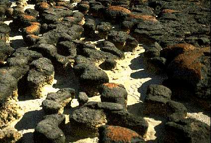
CYANOBACTERIA
General Characteristics
Cyanobacteria have a ubiquitous distribution with varied requirements and multiple appearances. Some produce STROMATOLITES when colonies bind calcium-rich sediments. They probably played a role in elevating the oxygen level in the early atmosphere. Many have a mucilagenous coating and a variety of colors. Some FIX nitrogen and are responsible for continuous production of rice on the same land. Some are symbiotic in protists and animals and function as chloroplasts under these conditions.
Shapes range from unicellular, to branched, to plates or colonial forms. They may form filaments and may exceed 1 meter in length.
Freshwater and marine forms contain gas vacuoles to regulate buoyancy. Under extreme conditions they may float to surface and form "blooms". Some forms secrete chemicals toxic to other organisms such as fish.
Photosynthetic bacteria
- Cyanobacteria contains chlorophyll a, carotenoids, a blue pigment (phycocyanin) and generally a red pigment (phycoerythrin). Photosynthesis is identical to that in photosynthetic eukaryotes. Their main storage product is glycogen.
- Green sulfur bacteria use sulfur to take the place of oxygen in the photosynthetic reaction. They require hydrogen sulfide or similar substrate and the elemental sulfur may accumulate in the cells.
- Purple sulfur bacteria are similar to the previous group. They contain red and yellow carotenoids.
- Purple nonsulfur bacteria are similar to previous group except other compounds, including alcohols, fatty acids, and keto acids serve as electron (hydrogen) donors for photosynthesis. Halobacterium halobium contains bacteriorhodopsin which responds to violet wavelengths and is the only nongreen living unit capable of converting radiant energy into chemical energy through photosynthesis.
- Prochloron - contains chlorophylls a and b and carotenoids. It
lives only in association with colonial marine animals (ascidians or sea squirts) and may be an evolutionary link to the green algae.
Plant Diseases
Bacterial diseases contribute to a world-wide crop loss of roughly 12-15 % annually. Almost any type of plant can be affected by bacteria. Virtually all plant-pathogenic bacteria are bacilli, and almost all occur in their host plants as parasites. Symptoms vary considerably with the most common appearing as SPOTS of various sizes on stems, leaves, and fruits. Almost all such bacterial SPOTS are caused by two closely related genera, Pseudomonas and Xanthomonas.
Some of the most destructive diseases of plants are caused by bacteria and are characterized as blights, soft rots, and wilts.
BLIGHTS - characterized by rapidly developing necroses (dead, discolored areas) on stems, leaves, and flowers. Fire blight in apples and pears may result in the death of young trees within a single season. Causative pathogen is Erwinia amylovora.
SOFT ROTS - occur most commonly in fleshy storage organs of vegetables, such as potatoes or onions, or in fleshy fruits, such as tomatoes or eggplants. Causative pathogen is usually of the genus Erwinia.
WILTS - affect the vascular systems of herbaceous plants. Vessels of the xylem are invaded and bacteria multiply there. Water and nutrient flow is impeded and wilting and death occur. Portions of the vessel wall may be destroyed and vessel may rupture leading to bacterial spread to adjacent parenchyma. Alfalfa and bean plants are affected by Corynebacterium, squash and watermelons by Erwinia tracheiphila, cabbage and other crucifers by Xanthomonas campestris.
Agrobacterium tumifaciens causes a disease known as crown gall in a variety of plants.
Mycoplasmas are a group of bacteria that are the smallest known cells. They are procaryotic but lack a cell wall. They seem to represent bacteria that have been simplified in their structure during their evolution. Over 50 plant diseases affecting more than 200 plant species have been associated with mycoplasmas. Most are confined to the sieve tubes of the phloem.

