


Morphology Based Classification of Apicomplexa
Coccidiosis is a parasitic disease caused by intracellular protozoa in vertebrates with lesser species infecting invertebrates. The disease is of economic and medical importance affecting man, sheep, birds, cattle and many other animals.
In sheep, coccidiosis is caused by parasites of the genus Eimeria and is an important disease in the sheep industry; it is especially important in pre-weaned and recently weaned lambs. While nearly all animals are exposed to coccidia, they may not show obvious signs of disease. Infected animals can shed low numbers of oocyts in faeces for a long time presenting a source of the infection for the young.This condition, known as subclinical coccidiosis, has a significant impact on the economics of animal production causing a reduction in weight gain and feed efficiency and increased susceptibility to other diseases. Clinical coccidiosis results in even higher financial losses for producers because of medical treatment costs, more severe effect on growth performance and sometimes death losses.
Infected animals are characterised by diarrhoea, inappetence, dehydration, weight loss, weakness, anaemia, depression, rough hair coat and, in severe outbreaks, death. Faeces become diarrhoeic but do not always contain blood. In an outbreak of coccidiosis the figure of morbidity can vary between 10 % to 40 % but mortality is rarely more than 10 %
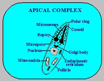
The coccidia of the suborder Eimeriina has been divided into two distinct groups: enteric coccidia (Eimeriidae and Cryptosporidiidae) and cysts-forming coccidia (Sarcocystiidae). Both the enteric coccidia and the cysts-forming coccidia species have great importance in the veterinary and medical fields. These parasites have been placed in the phylum Apicomplexa because of the presence of special organelles found in the parasites anterior end (the apical comlex plays a role in allowing parasites to get into host cells through an unknown mechanism).
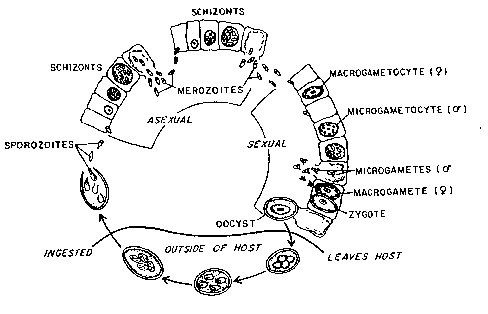
The life cycle of the family Eimeriidae consists of three principle processes:
i) Schizogony (asexual stage),
ii) Gametogony (sexual stage)
iii) Sporogony (asexual stage).
These three principle processes are completed in two different stages in the life cycle. One stage occurs in the intestine of the host (Endogenous stage) and at this stage schizogony and gametogony are completed in the host gut. The other stage occurs outside of the host (Exogenous stage) and this represents the third and last part of life cycle, sporogony
When the host ingests a sporulated oocyst the infection process begins and the host becomes infected. Under special stimuli such as an anaerobic environment in the presence of bile salts and enzymes, the sporozoites are released from the oocyst and sporocyst. The released sporozoites penetrate the cells of the intestine. Two types of penetration occur, one active the other passive; sporozoites either penetrate directly or are indirectly carried into the host cell by white blood cells.
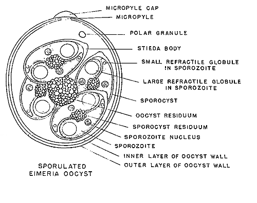
When sporozoites penetrate the host cell they change shape, becoming round (Trophozoites), increasing in number by mitosis (Shizogony or Merogony) to form first generation meronts. These first generation meronts break up the host cell walls and pass into the intestinal lumen. First generation meronts invade new host cells and again increase in number by mitosis forming second generation merozoites. When second generation meronts invade fresh host cells some develop into microgamonts (male gamonts, microgametocytes) while most develop into macrogamonts (macrogametocytes, female gamont). The sexual stage starts at this point and microgamonts increase in number as biflagellated microgametocytes. At the same time macrogametocytes grow in size several times. Microgametocytes burst out the host cell and when they reach a fresh cell which contains macrogametocytes fertilisation occurs. At this time wall forming bodies one and two (WFB1-WFB2) appears in the macrogametocytes. WFB1 is responsible for forming the outer layer of the oocysts wall and WFB2 forms the inner layer of the oocysts wall. After fertilisation, the wall forming bodies penetrate the cytoplasmic membrane and cover the macrogametocytes to form the oocyst wall. When the wall formation is complete, the macrogametocytes become an unsporulated fertile oocyst. These oocysts are released into the environment by faecal contamination
SEEP INFECTIVE EIMERIA SPECIES
TREATMENT:
Numerous drugs have been developed for the treatment of coccidiosis and prophylactic medication is preferred to therapeutic medication. For sheep coccidiosis, amprolium, monensin, lasalocid and decoquinate are the drugs of choice for prophylaxis while sulphonamides are used as chemotheraphetic agents despite being only partially effective. The ionophores, quinolones and clopidol act against the sporozoites and trophozoites while amprolium and sulphonamides act principally against later stages.
Good management reduces the risk of coccidia outbreaks in chickens and farm animals. For preventative medication several anticoccidials can be effective when given in food for 3 weeks before animals enter the feedlot and one week afterwards. Another stressful time for sheep is lambing time, ewes brought into confined lambing sheds saturate the environment with faeces containing oocysts, which accumulate in large numbers by the time lambs are born. Ewes should be given preventative medication at least one week before the beginning of lambing.


My Favorite Links
Veterinary Parasitology
Parasite and Parasitological Resource
Veterinary Parasitology II
Evolution and Phylogeny
Veterinary Medicine Drugs
Parasitology Test
Parasitology LAB Methods
eEmerging Infectious Disease
Epi6 Home Page
Dr.Metin Korkmaz Parasitology List
Livestock Breeds
